Iron »
PDB 4wha-4x33 »
4wvz »
Iron in PDB 4wvz: Crystal Structure of Artificial Crosslinked Thiol Dioxygenase G95C Variant From Pseudomonas Aeruginosa
Protein crystallography data
The structure of Crystal Structure of Artificial Crosslinked Thiol Dioxygenase G95C Variant From Pseudomonas Aeruginosa, PDB code: 4wvz
was solved by
M.Fellner,
E.P.Tchesnokov,
G.N.L.Jameson,
S.M.Wilbanks,
with X-Ray Crystallography technique. A brief refinement statistics is given in the table below:
| Resolution Low / High (Å) | 39.91 / 2.09 |
| Space group | P 41 21 2 |
| Cell size a, b, c (Å), α, β, γ (°) | 66.543, 66.543, 376.852, 90.00, 90.00, 90.00 |
| R / Rfree (%) | 17.5 / 21.9 |
Iron Binding Sites:
The binding sites of Iron atom in the Crystal Structure of Artificial Crosslinked Thiol Dioxygenase G95C Variant From Pseudomonas Aeruginosa
(pdb code 4wvz). This binding sites where shown within
5.0 Angstroms radius around Iron atom.
In total 4 binding sites of Iron where determined in the Crystal Structure of Artificial Crosslinked Thiol Dioxygenase G95C Variant From Pseudomonas Aeruginosa, PDB code: 4wvz:
Jump to Iron binding site number: 1; 2; 3; 4;
In total 4 binding sites of Iron where determined in the Crystal Structure of Artificial Crosslinked Thiol Dioxygenase G95C Variant From Pseudomonas Aeruginosa, PDB code: 4wvz:
Jump to Iron binding site number: 1; 2; 3; 4;
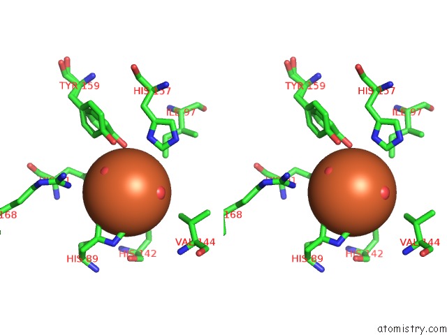
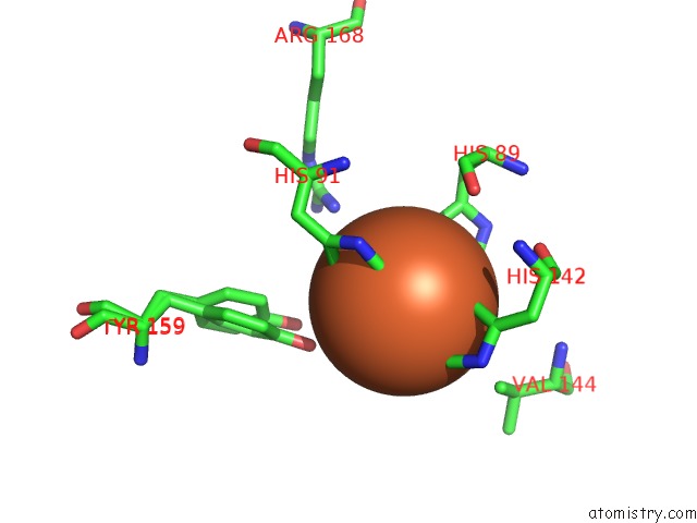
Iron binding site 1 out of 4 in 4wvz
Go back to
Iron binding site 1 out
of 4 in the Crystal Structure of Artificial Crosslinked Thiol Dioxygenase G95C Variant From Pseudomonas Aeruginosa
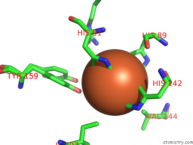
Mono view
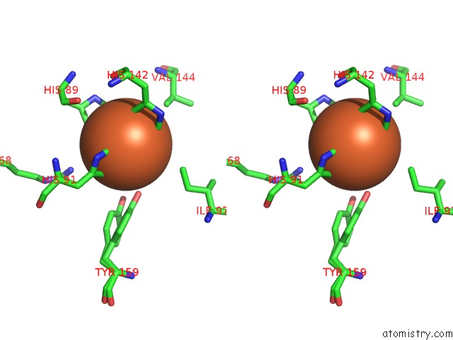
Stereo pair view
Mono view
Stereo pair view
A full contact list of Iron with other atoms in the Fe binding
site number 1 of Crystal Structure of Artificial Crosslinked Thiol Dioxygenase G95C Variant From Pseudomonas Aeruginosa within 5.0Å range:
|
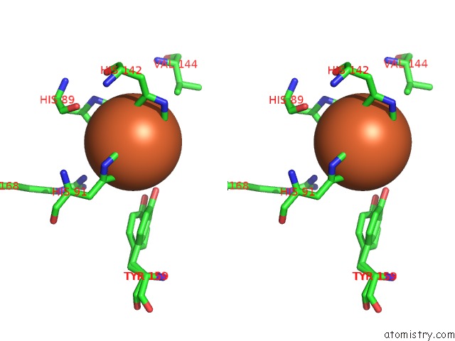
Iron binding site 2 out of 4 in 4wvz
Go back to
Iron binding site 2 out
of 4 in the Crystal Structure of Artificial Crosslinked Thiol Dioxygenase G95C Variant From Pseudomonas Aeruginosa
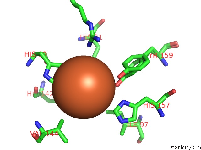
Mono view

Stereo pair view
Mono view
Stereo pair view
A full contact list of Iron with other atoms in the Fe binding
site number 2 of Crystal Structure of Artificial Crosslinked Thiol Dioxygenase G95C Variant From Pseudomonas Aeruginosa within 5.0Å range:
|
Iron binding site 3 out of 4 in 4wvz
Go back to
Iron binding site 3 out
of 4 in the Crystal Structure of Artificial Crosslinked Thiol Dioxygenase G95C Variant From Pseudomonas Aeruginosa
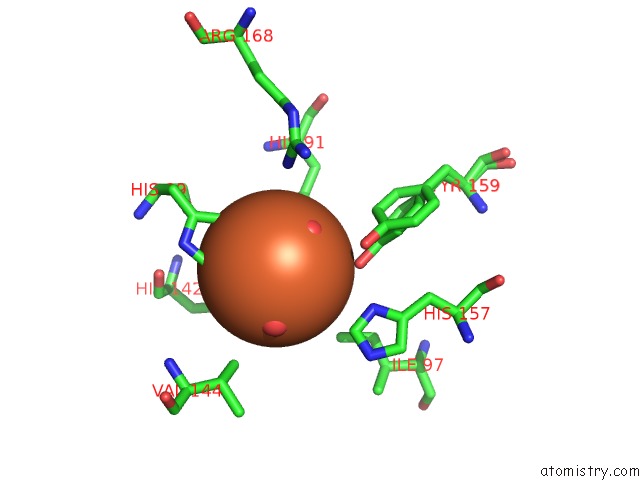
Mono view
Stereo pair view
Mono view
Stereo pair view
A full contact list of Iron with other atoms in the Fe binding
site number 3 of Crystal Structure of Artificial Crosslinked Thiol Dioxygenase G95C Variant From Pseudomonas Aeruginosa within 5.0Å range:
|
Iron binding site 4 out of 4 in 4wvz
Go back to
Iron binding site 4 out
of 4 in the Crystal Structure of Artificial Crosslinked Thiol Dioxygenase G95C Variant From Pseudomonas Aeruginosa
Mono view
Stereo pair view
Mono view
Stereo pair view
A full contact list of Iron with other atoms in the Fe binding
site number 4 of Crystal Structure of Artificial Crosslinked Thiol Dioxygenase G95C Variant From Pseudomonas Aeruginosa within 5.0Å range:
|
Reference:
M.Fellner,
S.Aloi,
E.P.Tchesnokov,
S.M.Wilbanks,
G.N.Jameson.
Substrate and pH-Dependent Kinetic Profile of 3-Mercaptopropionate Dioxygenase From Pseudomonas Aeruginosa. Biochemistry V. 55 1362 2016.
ISSN: ISSN 0006-2960
PubMed: 26878277
DOI: 10.1021/ACS.BIOCHEM.5B01203
Page generated: Mon Aug 5 15:00:01 2024
ISSN: ISSN 0006-2960
PubMed: 26878277
DOI: 10.1021/ACS.BIOCHEM.5B01203
Last articles
Fe in 2YXOFe in 2YRS
Fe in 2YXC
Fe in 2YNM
Fe in 2YVJ
Fe in 2YP1
Fe in 2YU2
Fe in 2YU1
Fe in 2YQB
Fe in 2YOO