Iron »
PDB 4x3s-4xq1 »
4xdp »
Iron in PDB 4xdp: Crystal Structure of Human KDM4C Catalytic Domain Bound to Tris
Protein crystallography data
The structure of Crystal Structure of Human KDM4C Catalytic Domain Bound to Tris, PDB code: 4xdp
was solved by
K.K.Swinger,
P.A.Boriack-Sjodin,
with X-Ray Crystallography technique. A brief refinement statistics is given in the table below:
| Resolution Low / High (Å) | 31.42 / 2.07 |
| Space group | C 1 2 1 |
| Cell size a, b, c (Å), α, β, γ (°) | 93.870, 89.750, 98.280, 90.00, 96.31, 90.00 |
| R / Rfree (%) | 20.7 / 24.2 |
Other elements in 4xdp:
The structure of Crystal Structure of Human KDM4C Catalytic Domain Bound to Tris also contains other interesting chemical elements:
| Chlorine | (Cl) | 6 atoms |
| Zinc | (Zn) | 2 atoms |
Iron Binding Sites:
The binding sites of Iron atom in the Crystal Structure of Human KDM4C Catalytic Domain Bound to Tris
(pdb code 4xdp). This binding sites where shown within
5.0 Angstroms radius around Iron atom.
In total 2 binding sites of Iron where determined in the Crystal Structure of Human KDM4C Catalytic Domain Bound to Tris, PDB code: 4xdp:
Jump to Iron binding site number: 1; 2;
In total 2 binding sites of Iron where determined in the Crystal Structure of Human KDM4C Catalytic Domain Bound to Tris, PDB code: 4xdp:
Jump to Iron binding site number: 1; 2;
Iron binding site 1 out of 2 in 4xdp
Go back to
Iron binding site 1 out
of 2 in the Crystal Structure of Human KDM4C Catalytic Domain Bound to Tris
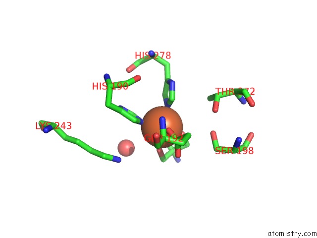
Mono view
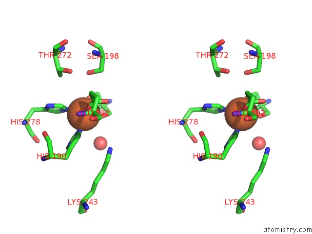
Stereo pair view
Mono view
Stereo pair view
A full contact list of Iron with other atoms in the Fe binding
site number 1 of Crystal Structure of Human KDM4C Catalytic Domain Bound to Tris within 5.0Å range:
|
Iron binding site 2 out of 2 in 4xdp
Go back to
Iron binding site 2 out
of 2 in the Crystal Structure of Human KDM4C Catalytic Domain Bound to Tris
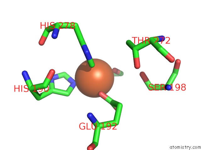
Mono view
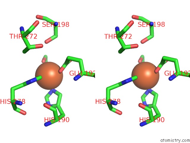
Stereo pair view
Mono view
Stereo pair view
A full contact list of Iron with other atoms in the Fe binding
site number 2 of Crystal Structure of Human KDM4C Catalytic Domain Bound to Tris within 5.0Å range:
|
Reference:
T.J.Wigle,
K.K.Swinger,
J.E.Campbell,
M.D.Scholle,
J.Sherrill,
E.A.Admirand,
P.A.Boriack-Sjodin,
K.W.Kuntz,
R.Chesworth,
M.P.Moyer,
M.P.Scott,
R.A.Copeland.
A High-Throughput Mass Spectrometry Assay Coupled with Redox Activity Testing Reduces Artifacts and False Positives in Lysine Demethylase Screening. J Biomol Screen 2015.
ISSN: ESSN 1552-454X
PubMed: 25755264
DOI: 10.1177/1087057115575689
Page generated: Mon Aug 5 15:29:38 2024
ISSN: ESSN 1552-454X
PubMed: 25755264
DOI: 10.1177/1087057115575689
Last articles
Cl in 7ZBACl in 7Z53
Cl in 7ZB9
Cl in 7Z93
Cl in 7Z88
Cl in 7Z8Y
Cl in 7Z8W
Cl in 7Z87
Cl in 7Z7B
Cl in 7Z5N