Iron »
PDB 4x3s-4xq1 »
4xmh »
Iron in PDB 4xmh: Crystal Structure of Nitrophorin 7 From Rhodnius Prolixus at pH 7.8 Complexed with Gly-Gly-Gly
Enzymatic activity of Crystal Structure of Nitrophorin 7 From Rhodnius Prolixus at pH 7.8 Complexed with Gly-Gly-Gly
All present enzymatic activity of Crystal Structure of Nitrophorin 7 From Rhodnius Prolixus at pH 7.8 Complexed with Gly-Gly-Gly:
1.7.6.1;
1.7.6.1;
Protein crystallography data
The structure of Crystal Structure of Nitrophorin 7 From Rhodnius Prolixus at pH 7.8 Complexed with Gly-Gly-Gly, PDB code: 4xmh
was solved by
H.Ogata,
with X-Ray Crystallography technique. A brief refinement statistics is given in the table below:
| Resolution Low / High (Å) | 34.71 / 1.29 |
| Space group | P 1 21 1 |
| Cell size a, b, c (Å), α, β, γ (°) | 38.374, 66.929, 38.885, 90.00, 116.79, 90.00 |
| R / Rfree (%) | 15.5 / 18.2 |
Iron Binding Sites:
The binding sites of Iron atom in the Crystal Structure of Nitrophorin 7 From Rhodnius Prolixus at pH 7.8 Complexed with Gly-Gly-Gly
(pdb code 4xmh). This binding sites where shown within
5.0 Angstroms radius around Iron atom.
In total only one binding site of Iron was determined in the Crystal Structure of Nitrophorin 7 From Rhodnius Prolixus at pH 7.8 Complexed with Gly-Gly-Gly, PDB code: 4xmh:
In total only one binding site of Iron was determined in the Crystal Structure of Nitrophorin 7 From Rhodnius Prolixus at pH 7.8 Complexed with Gly-Gly-Gly, PDB code: 4xmh:
Iron binding site 1 out of 1 in 4xmh
Go back to
Iron binding site 1 out
of 1 in the Crystal Structure of Nitrophorin 7 From Rhodnius Prolixus at pH 7.8 Complexed with Gly-Gly-Gly
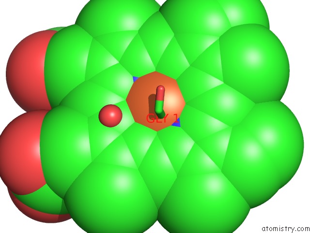
Mono view
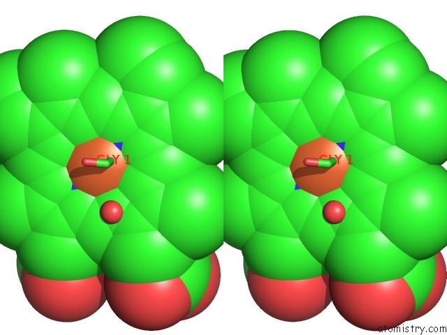
Stereo pair view
Mono view
Stereo pair view
A full contact list of Iron with other atoms in the Fe binding
site number 1 of Crystal Structure of Nitrophorin 7 From Rhodnius Prolixus at pH 7.8 Complexed with Gly-Gly-Gly within 5.0Å range:
|
Reference:
M.Knipp,
H.Ogata,
G.Soavi,
G.Cerullo,
A.Allegri,
S.Abbruzzetti,
S.Bruno,
C.Viappiani,
A.Bidon-Chanal,
F.J.Luque.
Structure and Dynamics of the Membrane Attaching Nitric Oxide Transporter Nitrophorin 7. F1000RES V. 4 45 2015.
ISSN: ISSN 2046-1402
PubMed: 26167269
DOI: 10.12688/F1000RESEARCH.6060.1
Page generated: Tue Aug 5 17:11:45 2025
ISSN: ISSN 2046-1402
PubMed: 26167269
DOI: 10.12688/F1000RESEARCH.6060.1
Last articles
Fe in 5AA5Fe in 5ADE
Fe in 5ADD
Fe in 5ADC
Fe in 5ADB
Fe in 5ADA
Fe in 5AD9
Fe in 5AD8
Fe in 5AD7
Fe in 5AD5