Iron »
PDB 4x3s-4xq1 »
4xpw »
Iron in PDB 4xpw: Crystal Structures of LEU114F Mutant
Protein crystallography data
The structure of Crystal Structures of LEU114F Mutant, PDB code: 4xpw
was solved by
P.Chuankhayan,
K.H.C.Chen,
H.H.Wu,
C.J.Chen,
M.Fukuda,
S.S.F.Yu,
S.I.Chan,
with X-Ray Crystallography technique. A brief refinement statistics is given in the table below:
| Resolution Low / High (Å) | 24.84 / 1.17 |
| Space group | P 6 |
| Cell size a, b, c (Å), α, β, γ (°) | 83.281, 83.281, 30.959, 90.00, 90.00, 120.00 |
| R / Rfree (%) | 15.2 / 16.5 |
Iron Binding Sites:
The binding sites of Iron atom in the Crystal Structures of LEU114F Mutant
(pdb code 4xpw). This binding sites where shown within
5.0 Angstroms radius around Iron atom.
In total 2 binding sites of Iron where determined in the Crystal Structures of LEU114F Mutant, PDB code: 4xpw:
Jump to Iron binding site number: 1; 2;
In total 2 binding sites of Iron where determined in the Crystal Structures of LEU114F Mutant, PDB code: 4xpw:
Jump to Iron binding site number: 1; 2;
Iron binding site 1 out of 2 in 4xpw
Go back to
Iron binding site 1 out
of 2 in the Crystal Structures of LEU114F Mutant
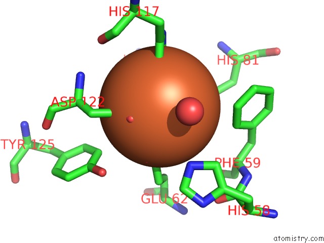
Mono view
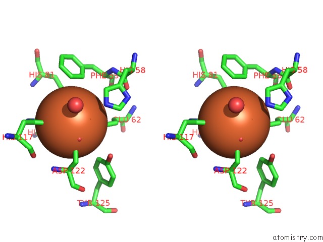
Stereo pair view
Mono view
Stereo pair view
A full contact list of Iron with other atoms in the Fe binding
site number 1 of Crystal Structures of LEU114F Mutant within 5.0Å range:
|
Iron binding site 2 out of 2 in 4xpw
Go back to
Iron binding site 2 out
of 2 in the Crystal Structures of LEU114F Mutant
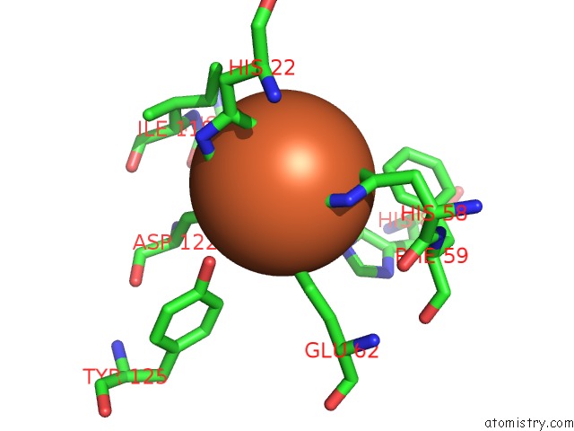
Mono view
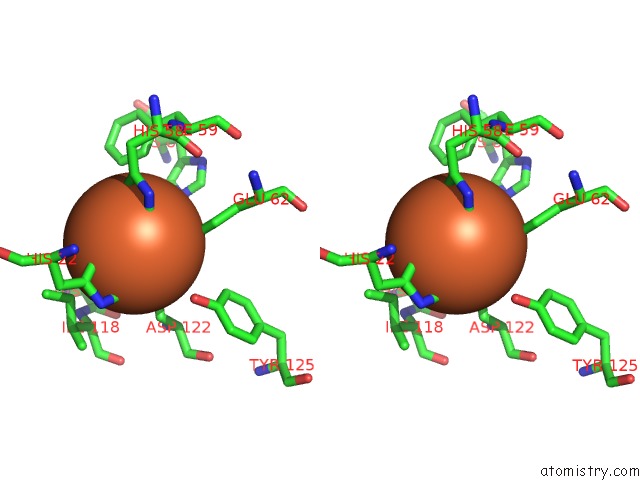
Stereo pair view
Mono view
Stereo pair view
A full contact list of Iron with other atoms in the Fe binding
site number 2 of Crystal Structures of LEU114F Mutant within 5.0Å range:
|
Reference:
K.H.Chen,
P.Chuankhayan,
H.H.Wu,
C.J.Chen,
M.Fukuda,
S.S.Yu,
S.I.Chan.
The Bacteriohemerythrin From Methylococcus Capsulatus (Bath): Crystal Structures Reveal That LEU114 Regulates A Water Tunnel. J.Inorg.Biochem. 2015.
ISSN: ISSN 0162-0134
PubMed: 25890483
DOI: 10.1016/J.JINORGBIO.2015.04.001
Page generated: Tue Aug 5 17:16:05 2025
ISSN: ISSN 0162-0134
PubMed: 25890483
DOI: 10.1016/J.JINORGBIO.2015.04.001
Last articles
Fe in 5B84Fe in 5B82
Fe in 5B72
Fe in 5B6Q
Fe in 5B50
Fe in 5B4F
Fe in 5B51
Fe in 5B48
Fe in 5B4E
Fe in 5B4Z