Iron »
PDB 4xry-4yoq »
4xva »
Iron in PDB 4xva: Crystal Structure of Wild Type Cytochrome C Peroxidase
Enzymatic activity of Crystal Structure of Wild Type Cytochrome C Peroxidase
All present enzymatic activity of Crystal Structure of Wild Type Cytochrome C Peroxidase:
1.11.1.5;
1.11.1.5;
Protein crystallography data
The structure of Crystal Structure of Wild Type Cytochrome C Peroxidase, PDB code: 4xva
was solved by
M.Fischer,
J.S.Fraser,
with X-Ray Crystallography technique. A brief refinement statistics is given in the table below:
| Resolution Low / High (Å) | 92.63 / 2.66 |
| Space group | P 21 21 21 |
| Cell size a, b, c (Å), α, β, γ (°) | 84.110, 104.760, 185.250, 90.00, 90.00, 90.00 |
| R / Rfree (%) | 23.3 / 27.6 |
Iron Binding Sites:
The binding sites of Iron atom in the Crystal Structure of Wild Type Cytochrome C Peroxidase
(pdb code 4xva). This binding sites where shown within
5.0 Angstroms radius around Iron atom.
In total 4 binding sites of Iron where determined in the Crystal Structure of Wild Type Cytochrome C Peroxidase, PDB code: 4xva:
Jump to Iron binding site number: 1; 2; 3; 4;
In total 4 binding sites of Iron where determined in the Crystal Structure of Wild Type Cytochrome C Peroxidase, PDB code: 4xva:
Jump to Iron binding site number: 1; 2; 3; 4;
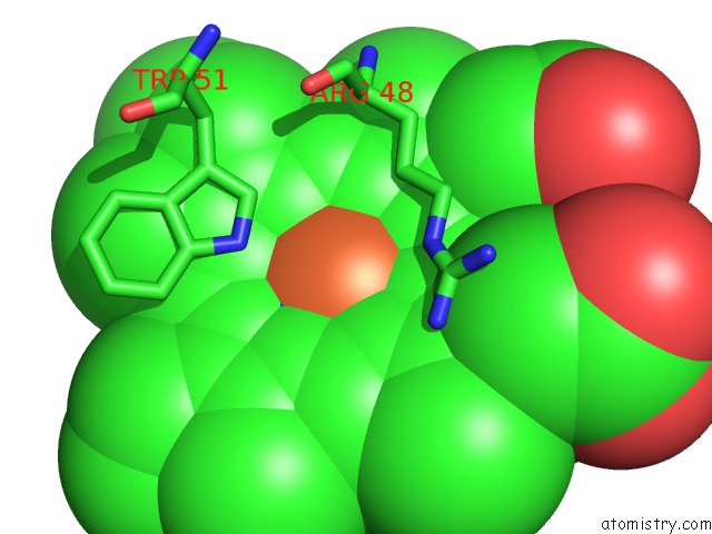
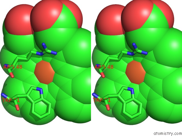
Iron binding site 1 out of 4 in 4xva
Go back to
Iron binding site 1 out
of 4 in the Crystal Structure of Wild Type Cytochrome C Peroxidase
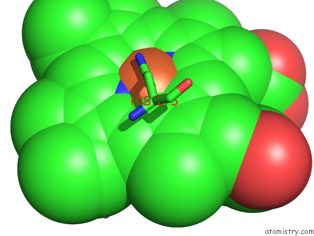
Mono view
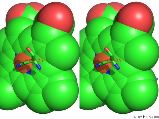
Stereo pair view
Mono view
Stereo pair view
A full contact list of Iron with other atoms in the Fe binding
site number 1 of Crystal Structure of Wild Type Cytochrome C Peroxidase within 5.0Å range:
|
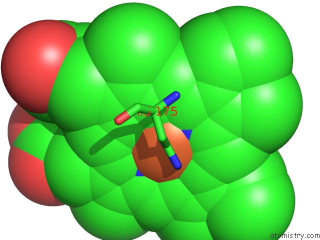
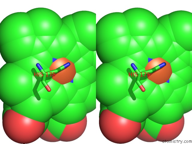
Iron binding site 2 out of 4 in 4xva
Go back to
Iron binding site 2 out
of 4 in the Crystal Structure of Wild Type Cytochrome C Peroxidase
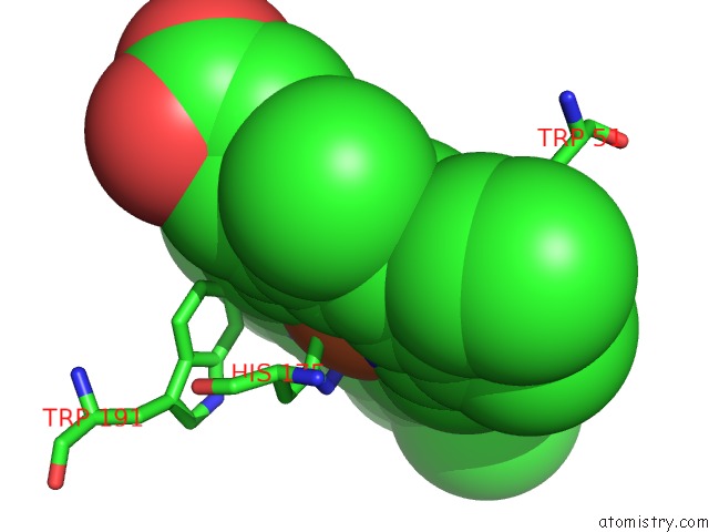
Mono view
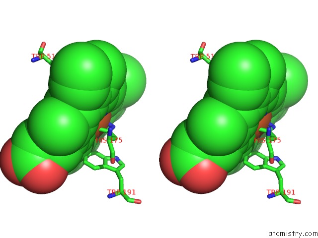
Stereo pair view
Mono view
Stereo pair view
A full contact list of Iron with other atoms in the Fe binding
site number 2 of Crystal Structure of Wild Type Cytochrome C Peroxidase within 5.0Å range:
|
Iron binding site 3 out of 4 in 4xva
Go back to
Iron binding site 3 out
of 4 in the Crystal Structure of Wild Type Cytochrome C Peroxidase
Mono view
Stereo pair view
Mono view
Stereo pair view
A full contact list of Iron with other atoms in the Fe binding
site number 3 of Crystal Structure of Wild Type Cytochrome C Peroxidase within 5.0Å range:
|
Iron binding site 4 out of 4 in 4xva
Go back to
Iron binding site 4 out
of 4 in the Crystal Structure of Wild Type Cytochrome C Peroxidase
Mono view
Stereo pair view
Mono view
Stereo pair view
A full contact list of Iron with other atoms in the Fe binding
site number 4 of Crystal Structure of Wild Type Cytochrome C Peroxidase within 5.0Å range:
|
Reference:
M.Fischer,
B.K.Shoichet,
J.S.Fraser.
Opposing Penalties For Fragment Binding Revealed By Multi-Temperature X-Ray Crystallography. To Be Published.
Page generated: Mon Aug 5 16:00:52 2024
Last articles
Fe in 2YXOFe in 2YRS
Fe in 2YXC
Fe in 2YNM
Fe in 2YVJ
Fe in 2YP1
Fe in 2YU2
Fe in 2YU1
Fe in 2YQB
Fe in 2YOO