Iron »
PDB 4xry-4yoq »
4y4s »
Iron in PDB 4y4s: Crystal Structure of Y75A Hasa Dimer From Yersinia Pseudotuberculosis
Protein crystallography data
The structure of Crystal Structure of Y75A Hasa Dimer From Yersinia Pseudotuberculosis, PDB code: 4y4s
was solved by
T.Hino,
M.Kanadani,
T.Muroki,
Y.Ishimaru,
Y.Wada,
T.Sato,
S.Ozaki,
with X-Ray Crystallography technique. A brief refinement statistics is given in the table below:
| Resolution Low / High (Å) | 32.95 / 1.75 |
| Space group | P 43 21 2 |
| Cell size a, b, c (Å), α, β, γ (°) | 52.760, 52.760, 140.570, 90.00, 90.00, 90.00 |
| R / Rfree (%) | 20.3 / 24 |
Iron Binding Sites:
The binding sites of Iron atom in the Crystal Structure of Y75A Hasa Dimer From Yersinia Pseudotuberculosis
(pdb code 4y4s). This binding sites where shown within
5.0 Angstroms radius around Iron atom.
In total only one binding site of Iron was determined in the Crystal Structure of Y75A Hasa Dimer From Yersinia Pseudotuberculosis, PDB code: 4y4s:
In total only one binding site of Iron was determined in the Crystal Structure of Y75A Hasa Dimer From Yersinia Pseudotuberculosis, PDB code: 4y4s:
Iron binding site 1 out of 1 in 4y4s
Go back to
Iron binding site 1 out
of 1 in the Crystal Structure of Y75A Hasa Dimer From Yersinia Pseudotuberculosis
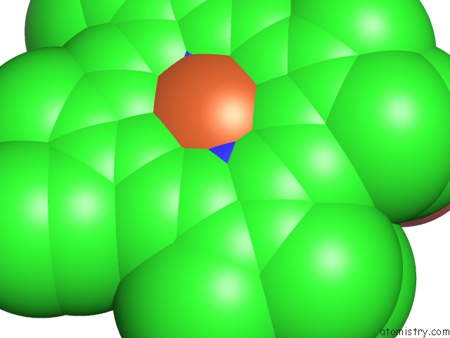
Mono view
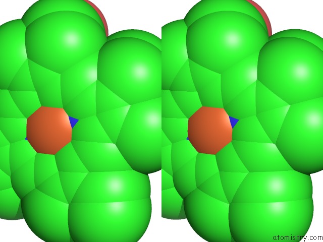
Stereo pair view
Mono view
Stereo pair view
A full contact list of Iron with other atoms in the Fe binding
site number 1 of Crystal Structure of Y75A Hasa Dimer From Yersinia Pseudotuberculosis within 5.0Å range:
|
Reference:
M.Kanadani,
T.Sato,
T.Hino,
S.Nagano,
S.Ozaki.
The Crystal Structure of Heme Acquisition System A From Yersinia Pseudotuberculosis (Hasaypt): Roles of the Axial Ligand TYR75 and Two Distal Arginines in Heme Binding J.Inorg.Biochem. V. 151 26 2015.
ISSN: ISSN 0162-0134
PubMed: 26210321
DOI: 10.1016/J.JINORGBIO.2015.07.007
Page generated: Mon Aug 5 16:05:46 2024
ISSN: ISSN 0162-0134
PubMed: 26210321
DOI: 10.1016/J.JINORGBIO.2015.07.007
Last articles
Zn in 9MJ5Zn in 9HNW
Zn in 9G0L
Zn in 9FNE
Zn in 9DZN
Zn in 9E0I
Zn in 9D32
Zn in 9DAK
Zn in 8ZXC
Zn in 8ZUF