Iron »
PDB 4yph-4z6p »
4z6m »
Iron in PDB 4z6m: Structure of H200Q Variant of Homoprotocatechuate 2,3-Dioxygenase From B.Fuscum at 1.35 Ang Resolution
Protein crystallography data
The structure of Structure of H200Q Variant of Homoprotocatechuate 2,3-Dioxygenase From B.Fuscum at 1.35 Ang Resolution, PDB code: 4z6m
was solved by
E.G.Kovaleva,
J.D.Lipscomb,
with X-Ray Crystallography technique. A brief refinement statistics is given in the table below:
| Resolution Low / High (Å) | 48.09 / 1.35 |
| Space group | P 21 21 2 |
| Cell size a, b, c (Å), α, β, γ (°) | 110.485, 152.089, 96.188, 90.00, 90.00, 90.00 |
| R / Rfree (%) | 11.2 / 14 |
Other elements in 4z6m:
The structure of Structure of H200Q Variant of Homoprotocatechuate 2,3-Dioxygenase From B.Fuscum at 1.35 Ang Resolution also contains other interesting chemical elements:
| Chlorine | (Cl) | 4 atoms |
| Calcium | (Ca) | 1 atom |
Iron Binding Sites:
The binding sites of Iron atom in the Structure of H200Q Variant of Homoprotocatechuate 2,3-Dioxygenase From B.Fuscum at 1.35 Ang Resolution
(pdb code 4z6m). This binding sites where shown within
5.0 Angstroms radius around Iron atom.
In total 4 binding sites of Iron where determined in the Structure of H200Q Variant of Homoprotocatechuate 2,3-Dioxygenase From B.Fuscum at 1.35 Ang Resolution, PDB code: 4z6m:
Jump to Iron binding site number: 1; 2; 3; 4;
In total 4 binding sites of Iron where determined in the Structure of H200Q Variant of Homoprotocatechuate 2,3-Dioxygenase From B.Fuscum at 1.35 Ang Resolution, PDB code: 4z6m:
Jump to Iron binding site number: 1; 2; 3; 4;
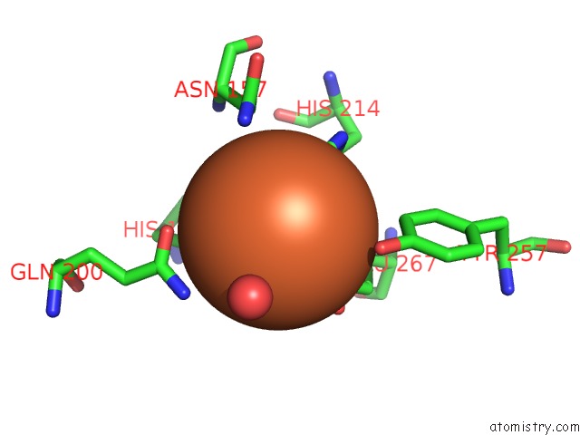
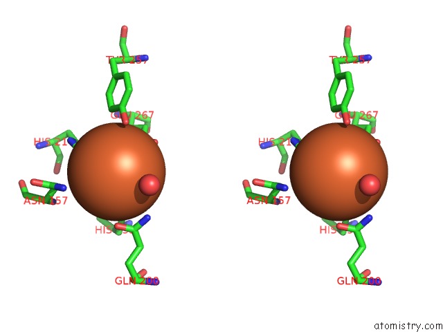
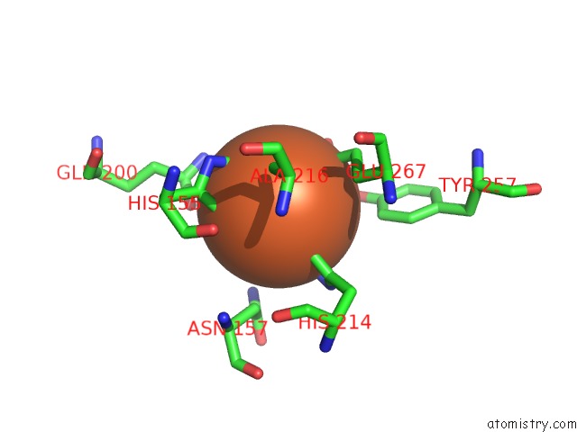
Iron binding site 1 out of 4 in 4z6m
Go back to
Iron binding site 1 out
of 4 in the Structure of H200Q Variant of Homoprotocatechuate 2,3-Dioxygenase From B.Fuscum at 1.35 Ang Resolution
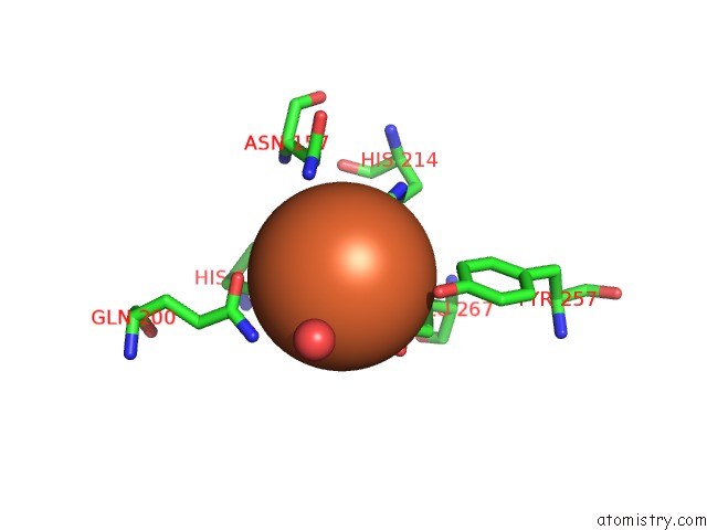
Mono view
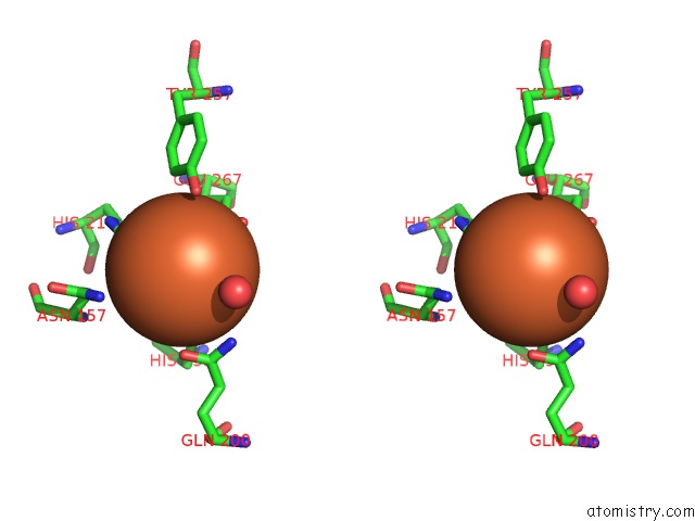
Stereo pair view
Mono view
Stereo pair view
A full contact list of Iron with other atoms in the Fe binding
site number 1 of Structure of H200Q Variant of Homoprotocatechuate 2,3-Dioxygenase From B.Fuscum at 1.35 Ang Resolution within 5.0Å range:
|
Iron binding site 2 out of 4 in 4z6m
Go back to
Iron binding site 2 out
of 4 in the Structure of H200Q Variant of Homoprotocatechuate 2,3-Dioxygenase From B.Fuscum at 1.35 Ang Resolution
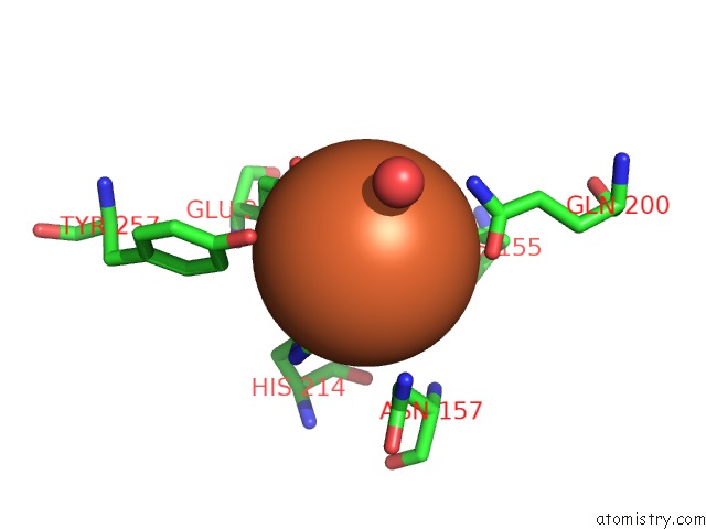
Mono view
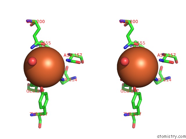
Stereo pair view
Mono view
Stereo pair view
A full contact list of Iron with other atoms in the Fe binding
site number 2 of Structure of H200Q Variant of Homoprotocatechuate 2,3-Dioxygenase From B.Fuscum at 1.35 Ang Resolution within 5.0Å range:
|
Iron binding site 3 out of 4 in 4z6m
Go back to
Iron binding site 3 out
of 4 in the Structure of H200Q Variant of Homoprotocatechuate 2,3-Dioxygenase From B.Fuscum at 1.35 Ang Resolution
Mono view
Stereo pair view
Mono view
Stereo pair view
A full contact list of Iron with other atoms in the Fe binding
site number 3 of Structure of H200Q Variant of Homoprotocatechuate 2,3-Dioxygenase From B.Fuscum at 1.35 Ang Resolution within 5.0Å range:
|
Iron binding site 4 out of 4 in 4z6m
Go back to
Iron binding site 4 out
of 4 in the Structure of H200Q Variant of Homoprotocatechuate 2,3-Dioxygenase From B.Fuscum at 1.35 Ang Resolution
Mono view

Stereo pair view
Mono view
Stereo pair view
A full contact list of Iron with other atoms in the Fe binding
site number 4 of Structure of H200Q Variant of Homoprotocatechuate 2,3-Dioxygenase From B.Fuscum at 1.35 Ang Resolution within 5.0Å range:
|
Reference:
E.G.Kovaleva,
M.S.Rogers,
J.D.Lipscomb.
Structural Basis For Substrate and Oxygen Activation in Homoprotocatechuate 2,3-Dioxygenase: Roles of Conserved Active Site Histidine 200. Biochemistry V. 54 5329 2015.
ISSN: ISSN 0006-2960
PubMed: 26267790
DOI: 10.1021/ACS.BIOCHEM.5B00709
Page generated: Mon Aug 5 17:11:57 2024
ISSN: ISSN 0006-2960
PubMed: 26267790
DOI: 10.1021/ACS.BIOCHEM.5B00709
Last articles
F in 7LY8F in 7LWG
F in 7LZV
F in 7LZF
F in 7LZD
F in 7LZA
F in 7LVX
F in 7LUN
F in 7LVR
F in 7LUK