Iron »
PDB 4z6q-4zn3 »
4z6q »
Iron in PDB 4z6q: Structure of H200N Variant of Homoprotocatechuate 2,3-Dioxygenase From B.Fuscum in Complex with Hpca at 1.57 Ang Resolution
Protein crystallography data
The structure of Structure of H200N Variant of Homoprotocatechuate 2,3-Dioxygenase From B.Fuscum in Complex with Hpca at 1.57 Ang Resolution, PDB code: 4z6q
was solved by
E.G.Kovaleva,
J.D.Lipscomb,
with X-Ray Crystallography technique. A brief refinement statistics is given in the table below:
| Resolution Low / High (Å) | 45.69 / 1.57 |
| Space group | P 21 21 2 |
| Cell size a, b, c (Å), α, β, γ (°) | 110.507, 150.077, 95.936, 90.00, 90.00, 90.00 |
| R / Rfree (%) | 14.1 / 16.5 |
Other elements in 4z6q:
The structure of Structure of H200N Variant of Homoprotocatechuate 2,3-Dioxygenase From B.Fuscum in Complex with Hpca at 1.57 Ang Resolution also contains other interesting chemical elements:
| Chlorine | (Cl) | 2 atoms |
| Calcium | (Ca) | 1 atom |
Iron Binding Sites:
The binding sites of Iron atom in the Structure of H200N Variant of Homoprotocatechuate 2,3-Dioxygenase From B.Fuscum in Complex with Hpca at 1.57 Ang Resolution
(pdb code 4z6q). This binding sites where shown within
5.0 Angstroms radius around Iron atom.
In total 4 binding sites of Iron where determined in the Structure of H200N Variant of Homoprotocatechuate 2,3-Dioxygenase From B.Fuscum in Complex with Hpca at 1.57 Ang Resolution, PDB code: 4z6q:
Jump to Iron binding site number: 1; 2; 3; 4;
In total 4 binding sites of Iron where determined in the Structure of H200N Variant of Homoprotocatechuate 2,3-Dioxygenase From B.Fuscum in Complex with Hpca at 1.57 Ang Resolution, PDB code: 4z6q:
Jump to Iron binding site number: 1; 2; 3; 4;
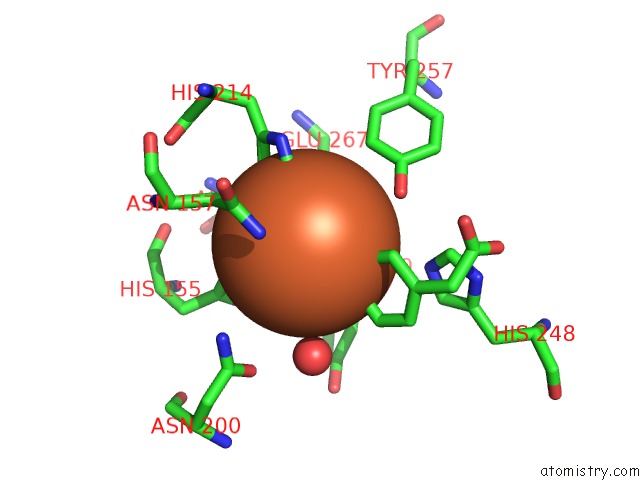
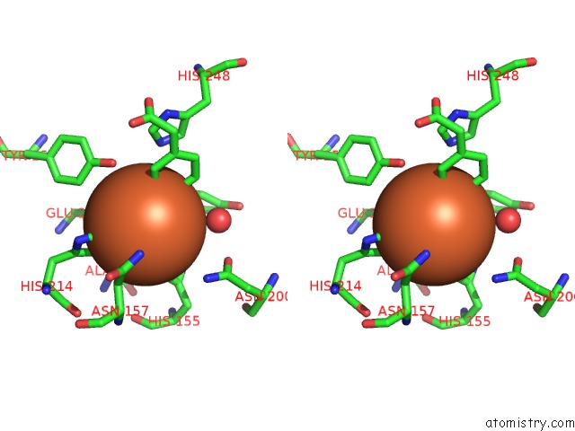
Iron binding site 1 out of 4 in 4z6q
Go back to
Iron binding site 1 out
of 4 in the Structure of H200N Variant of Homoprotocatechuate 2,3-Dioxygenase From B.Fuscum in Complex with Hpca at 1.57 Ang Resolution
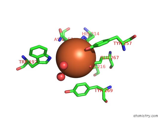
Mono view
Stereo pair view
Mono view
Stereo pair view
A full contact list of Iron with other atoms in the Fe binding
site number 1 of Structure of H200N Variant of Homoprotocatechuate 2,3-Dioxygenase From B.Fuscum in Complex with Hpca at 1.57 Ang Resolution within 5.0Å range:
|
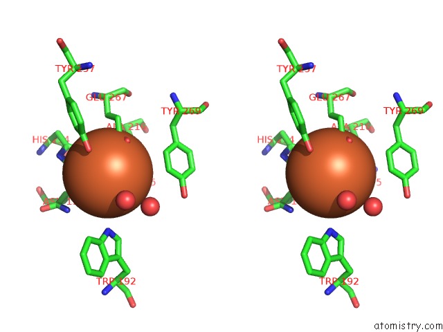
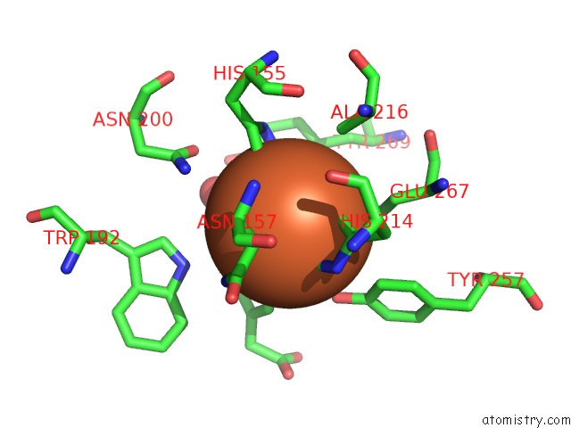
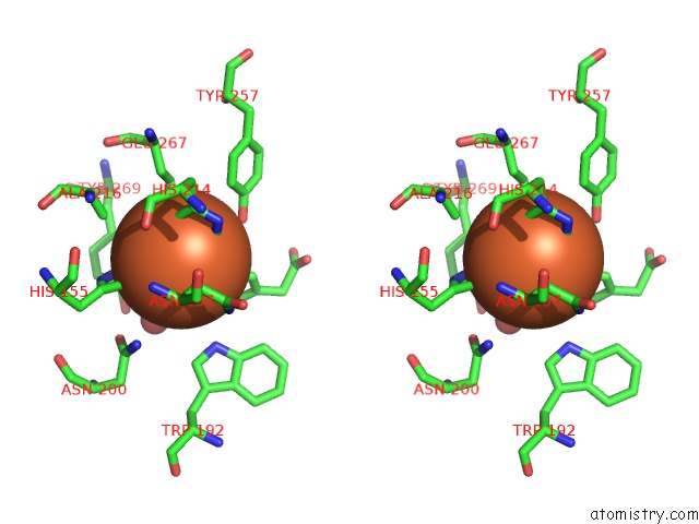
Iron binding site 2 out of 4 in 4z6q
Go back to
Iron binding site 2 out
of 4 in the Structure of H200N Variant of Homoprotocatechuate 2,3-Dioxygenase From B.Fuscum in Complex with Hpca at 1.57 Ang Resolution
Mono view
Stereo pair view
Mono view
Stereo pair view
A full contact list of Iron with other atoms in the Fe binding
site number 2 of Structure of H200N Variant of Homoprotocatechuate 2,3-Dioxygenase From B.Fuscum in Complex with Hpca at 1.57 Ang Resolution within 5.0Å range:
|
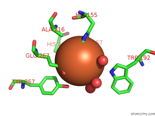
Iron binding site 3 out of 4 in 4z6q
Go back to
Iron binding site 3 out
of 4 in the Structure of H200N Variant of Homoprotocatechuate 2,3-Dioxygenase From B.Fuscum in Complex with Hpca at 1.57 Ang Resolution
Mono view
Stereo pair view
Mono view
Stereo pair view
A full contact list of Iron with other atoms in the Fe binding
site number 3 of Structure of H200N Variant of Homoprotocatechuate 2,3-Dioxygenase From B.Fuscum in Complex with Hpca at 1.57 Ang Resolution within 5.0Å range:
|
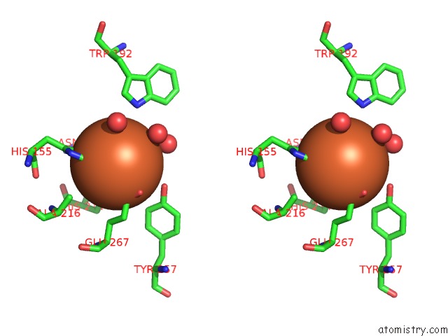
Iron binding site 4 out of 4 in 4z6q
Go back to
Iron binding site 4 out
of 4 in the Structure of H200N Variant of Homoprotocatechuate 2,3-Dioxygenase From B.Fuscum in Complex with Hpca at 1.57 Ang Resolution
Mono view
Stereo pair view
Mono view
Stereo pair view
A full contact list of Iron with other atoms in the Fe binding
site number 4 of Structure of H200N Variant of Homoprotocatechuate 2,3-Dioxygenase From B.Fuscum in Complex with Hpca at 1.57 Ang Resolution within 5.0Å range:
|
Reference:
E.G.Kovaleva,
M.S.Rogers,
J.D.Lipscomb.
Structural Basis For Substrate and Oxygen Activation in Homoprotocatechuate 2,3-Dioxygenase: Roles of Conserved Active Site Histidine 200. Biochemistry V. 54 5329 2015.
ISSN: ISSN 0006-2960
PubMed: 26267790
DOI: 10.1021/ACS.BIOCHEM.5B00709
Page generated: Mon Aug 5 18:05:53 2024
ISSN: ISSN 0006-2960
PubMed: 26267790
DOI: 10.1021/ACS.BIOCHEM.5B00709
Last articles
Zn in 9MJ5Zn in 9HNW
Zn in 9G0L
Zn in 9FNE
Zn in 9DZN
Zn in 9E0I
Zn in 9D32
Zn in 9DAK
Zn in 8ZXC
Zn in 8ZUF