Iron »
PDB 5adf-5az3 »
5ao4 »
Iron in PDB 5ao4: Crystal Structure of in Vitro Phosphorylated Human SAMHD1 (Amino Acid Residues 115-626) Bound to Gtp
Protein crystallography data
The structure of Crystal Structure of in Vitro Phosphorylated Human SAMHD1 (Amino Acid Residues 115-626) Bound to Gtp, PDB code: 5ao4
was solved by
L.H.Arnold,
D.Schwefel,
I.A.Taylor,
with X-Ray Crystallography technique. A brief refinement statistics is given in the table below:
| Resolution Low / High (Å) | 49.05 / 3.70 |
| Space group | C 1 2 1 |
| Cell size a, b, c (Å), α, β, γ (°) | 197.545, 80.817, 147.665, 90.00, 114.93, 90.00 |
| R / Rfree (%) | 24.4 / 30.8 |
Iron Binding Sites:
The binding sites of Iron atom in the Crystal Structure of in Vitro Phosphorylated Human SAMHD1 (Amino Acid Residues 115-626) Bound to Gtp
(pdb code 5ao4). This binding sites where shown within
5.0 Angstroms radius around Iron atom.
In total 4 binding sites of Iron where determined in the Crystal Structure of in Vitro Phosphorylated Human SAMHD1 (Amino Acid Residues 115-626) Bound to Gtp, PDB code: 5ao4:
Jump to Iron binding site number: 1; 2; 3; 4;
In total 4 binding sites of Iron where determined in the Crystal Structure of in Vitro Phosphorylated Human SAMHD1 (Amino Acid Residues 115-626) Bound to Gtp, PDB code: 5ao4:
Jump to Iron binding site number: 1; 2; 3; 4;
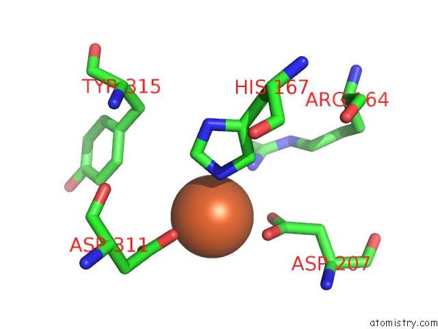
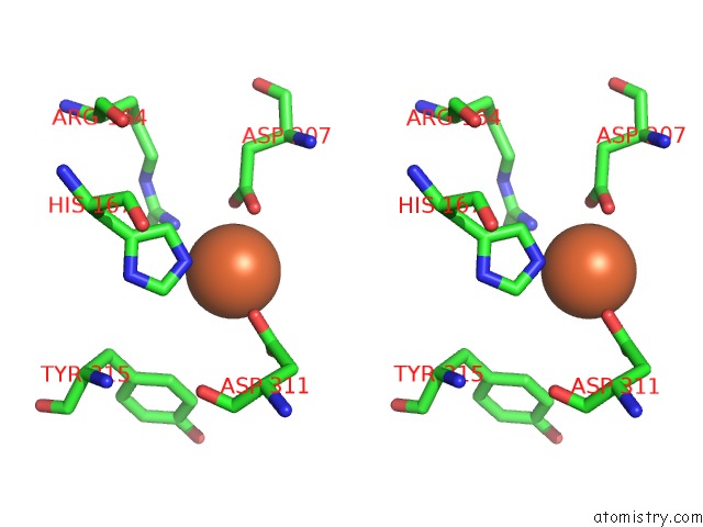
Iron binding site 1 out of 4 in 5ao4
Go back to
Iron binding site 1 out
of 4 in the Crystal Structure of in Vitro Phosphorylated Human SAMHD1 (Amino Acid Residues 115-626) Bound to Gtp
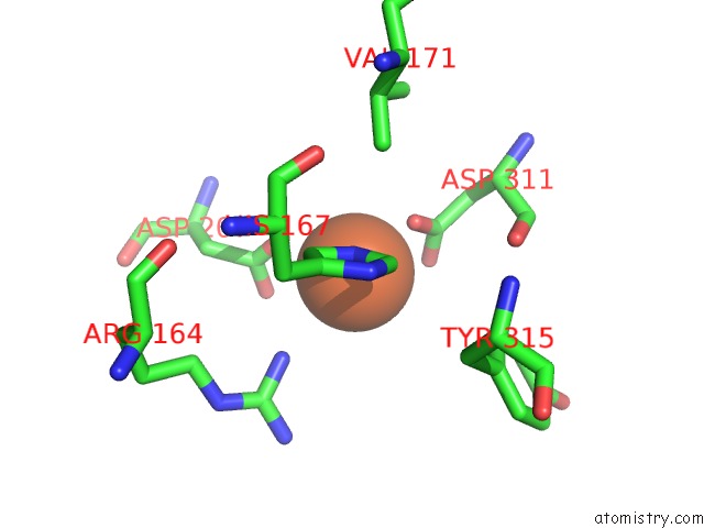
Mono view
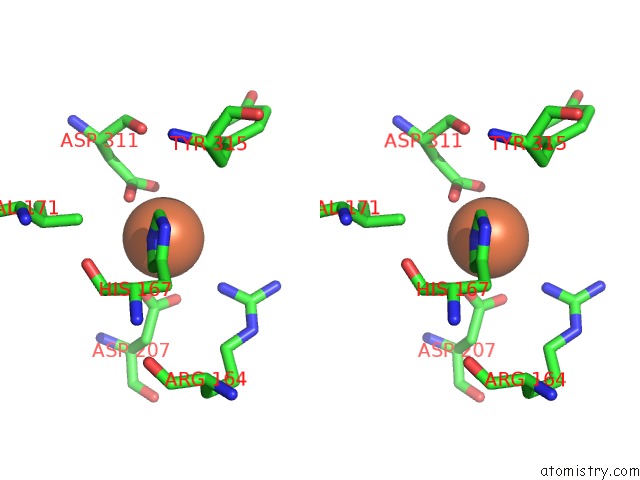
Stereo pair view
Mono view
Stereo pair view
A full contact list of Iron with other atoms in the Fe binding
site number 1 of Crystal Structure of in Vitro Phosphorylated Human SAMHD1 (Amino Acid Residues 115-626) Bound to Gtp within 5.0Å range:
|
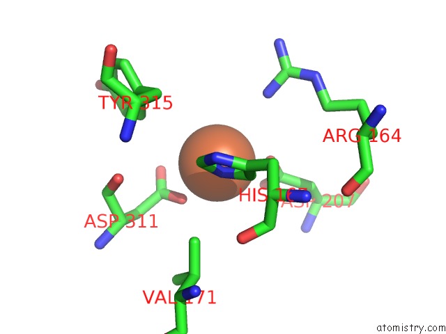
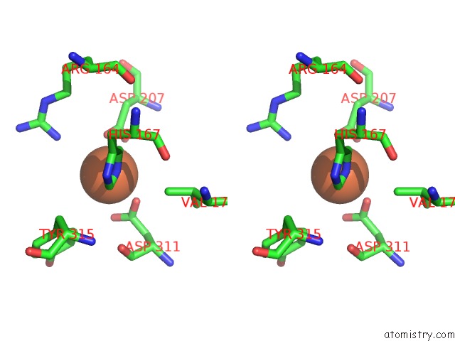
Iron binding site 2 out of 4 in 5ao4
Go back to
Iron binding site 2 out
of 4 in the Crystal Structure of in Vitro Phosphorylated Human SAMHD1 (Amino Acid Residues 115-626) Bound to Gtp
Mono view
Stereo pair view
Mono view
Stereo pair view
A full contact list of Iron with other atoms in the Fe binding
site number 2 of Crystal Structure of in Vitro Phosphorylated Human SAMHD1 (Amino Acid Residues 115-626) Bound to Gtp within 5.0Å range:
|
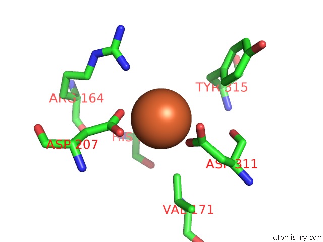
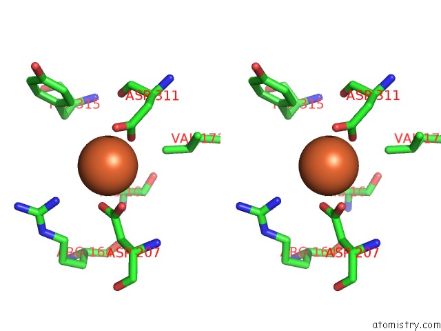
Iron binding site 3 out of 4 in 5ao4
Go back to
Iron binding site 3 out
of 4 in the Crystal Structure of in Vitro Phosphorylated Human SAMHD1 (Amino Acid Residues 115-626) Bound to Gtp
Mono view
Stereo pair view
Mono view
Stereo pair view
A full contact list of Iron with other atoms in the Fe binding
site number 3 of Crystal Structure of in Vitro Phosphorylated Human SAMHD1 (Amino Acid Residues 115-626) Bound to Gtp within 5.0Å range:
|
Iron binding site 4 out of 4 in 5ao4
Go back to
Iron binding site 4 out
of 4 in the Crystal Structure of in Vitro Phosphorylated Human SAMHD1 (Amino Acid Residues 115-626) Bound to Gtp
Mono view
Stereo pair view
Mono view
Stereo pair view
A full contact list of Iron with other atoms in the Fe binding
site number 4 of Crystal Structure of in Vitro Phosphorylated Human SAMHD1 (Amino Acid Residues 115-626) Bound to Gtp within 5.0Å range:
|
Reference:
L.H.Arnold,
H.C.T.Groom,
S.Kunzelmann,
D.Schwefel,
S.J.Caswell,
P.Ordonez,
M.C.Mann,
S.Rueschenbaum,
D.C.Goldstone,
S.Pennell,
S.A.Howell,
J.P.Stoye,
M.Webb,
I.A.Taylor,
K.N.Bishop.
Phospho-Dependent Regulation of SAMHD1 Oligomerisation Couples Catalysis and Restriction. Plos Pathog. V. 11 5194 2015.
ISSN: ISSN 1553-7366
PubMed: 26431200
DOI: 10.1371/JOURNAL.PPAT.1005194
Page generated: Tue Aug 5 19:06:46 2025
ISSN: ISSN 1553-7366
PubMed: 26431200
DOI: 10.1371/JOURNAL.PPAT.1005194
Last articles
Fe in 6N1FFe in 6N0K
Fe in 6N0J
Fe in 6N03
Fe in 6N02
Fe in 6MYS
Fe in 6MYR
Fe in 6MYQ
Fe in 6MSO
Fe in 6MYP