Iron »
PDB 5adf-5az3 »
5aui »
Iron in PDB 5aui: Crystal Structure of Ferredoxin
Protein crystallography data
The structure of Crystal Structure of Ferredoxin, PDB code: 5aui
was solved by
G.Kurisu,
K.Shinmura,
with X-Ray Crystallography technique. A brief refinement statistics is given in the table below:
| Resolution Low / High (Å) | 32.25 / 1.50 |
| Space group | C 1 2 1 |
| Cell size a, b, c (Å), α, β, γ (°) | 56.441, 53.314, 32.278, 90.00, 92.38, 90.00 |
| R / Rfree (%) | 17.2 / 19.3 |
Iron Binding Sites:
The binding sites of Iron atom in the Crystal Structure of Ferredoxin
(pdb code 5aui). This binding sites where shown within
5.0 Angstroms radius around Iron atom.
In total 2 binding sites of Iron where determined in the Crystal Structure of Ferredoxin, PDB code: 5aui:
Jump to Iron binding site number: 1; 2;
In total 2 binding sites of Iron where determined in the Crystal Structure of Ferredoxin, PDB code: 5aui:
Jump to Iron binding site number: 1; 2;
Iron binding site 1 out of 2 in 5aui
Go back to
Iron binding site 1 out
of 2 in the Crystal Structure of Ferredoxin
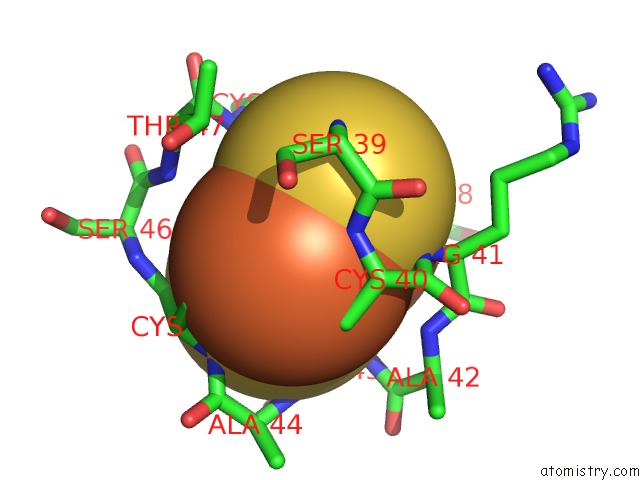
Mono view
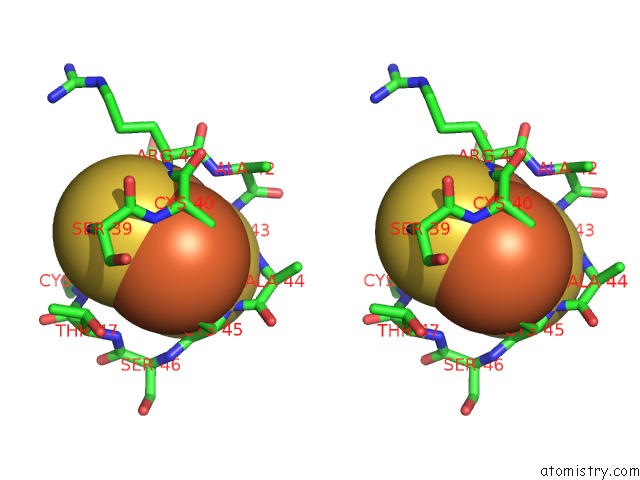
Stereo pair view
Mono view
Stereo pair view
A full contact list of Iron with other atoms in the Fe binding
site number 1 of Crystal Structure of Ferredoxin within 5.0Å range:
|
Iron binding site 2 out of 2 in 5aui
Go back to
Iron binding site 2 out
of 2 in the Crystal Structure of Ferredoxin
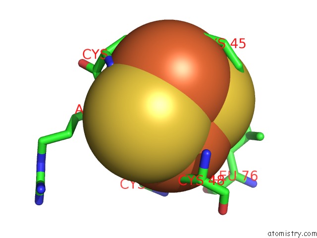
Mono view
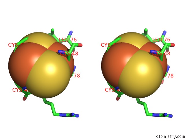
Stereo pair view
Mono view
Stereo pair view
A full contact list of Iron with other atoms in the Fe binding
site number 2 of Crystal Structure of Ferredoxin within 5.0Å range:
|
Reference:
R.Mutoh,
N.Muraki,
K.Shinmura,
H.Kubota-Kawai,
Y.H.Lee,
M.M.Nowaczyk,
M.Rogner,
T.Hase,
T.Ikegami,
G.Kurisu.
X-Ray Structure and Nuclear Magnetic Resonance Analysis of the Interaction Sites of the Ga-Substituted Cyanobacterial Ferredoxin Biochemistry V. 54 6052 2015.
ISSN: ISSN 0006-2960
PubMed: 26348494
DOI: 10.1021/ACS.BIOCHEM.5B00601
Page generated: Mon Aug 5 19:42:46 2024
ISSN: ISSN 0006-2960
PubMed: 26348494
DOI: 10.1021/ACS.BIOCHEM.5B00601
Last articles
Zn in 9JYWZn in 9IR4
Zn in 9IR3
Zn in 9GMX
Zn in 9GMW
Zn in 9JEJ
Zn in 9ERF
Zn in 9ERE
Zn in 9EGV
Zn in 9EGW