Iron »
PDB 5b1a-5c1v »
5b2x »
Iron in PDB 5b2x: Crystal Structure of P450BM3 Mutant with N-Perfluoroheptanoyl-L- Tryptophan
Enzymatic activity of Crystal Structure of P450BM3 Mutant with N-Perfluoroheptanoyl-L- Tryptophan
All present enzymatic activity of Crystal Structure of P450BM3 Mutant with N-Perfluoroheptanoyl-L- Tryptophan:
1.14.14.1; 1.6.2.4;
1.14.14.1; 1.6.2.4;
Protein crystallography data
The structure of Crystal Structure of P450BM3 Mutant with N-Perfluoroheptanoyl-L- Tryptophan, PDB code: 5b2x
was solved by
Z.Cong,
O.Shoji,
C.Kasai,
H.Sugimoto,
Y.Shiro,
Y.Watanabe,
with X-Ray Crystallography technique. A brief refinement statistics is given in the table below:
| Resolution Low / High (Å) | 19.17 / 1.90 |
| Space group | P 1 21 1 |
| Cell size a, b, c (Å), α, β, γ (°) | 58.402, 147.355, 64.301, 90.00, 100.10, 90.00 |
| R / Rfree (%) | 18 / 21.6 |
Other elements in 5b2x:
The structure of Crystal Structure of P450BM3 Mutant with N-Perfluoroheptanoyl-L- Tryptophan also contains other interesting chemical elements:
| Fluorine | (F) | 26 atoms |
Iron Binding Sites:
The binding sites of Iron atom in the Crystal Structure of P450BM3 Mutant with N-Perfluoroheptanoyl-L- Tryptophan
(pdb code 5b2x). This binding sites where shown within
5.0 Angstroms radius around Iron atom.
In total 2 binding sites of Iron where determined in the Crystal Structure of P450BM3 Mutant with N-Perfluoroheptanoyl-L- Tryptophan, PDB code: 5b2x:
Jump to Iron binding site number: 1; 2;
In total 2 binding sites of Iron where determined in the Crystal Structure of P450BM3 Mutant with N-Perfluoroheptanoyl-L- Tryptophan, PDB code: 5b2x:
Jump to Iron binding site number: 1; 2;
Iron binding site 1 out of 2 in 5b2x
Go back to
Iron binding site 1 out
of 2 in the Crystal Structure of P450BM3 Mutant with N-Perfluoroheptanoyl-L- Tryptophan
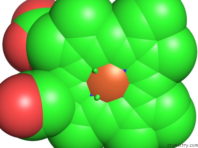
Mono view
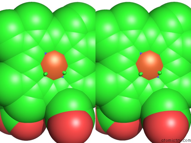
Stereo pair view
Mono view
Stereo pair view
A full contact list of Iron with other atoms in the Fe binding
site number 1 of Crystal Structure of P450BM3 Mutant with N-Perfluoroheptanoyl-L- Tryptophan within 5.0Å range:
|
Iron binding site 2 out of 2 in 5b2x
Go back to
Iron binding site 2 out
of 2 in the Crystal Structure of P450BM3 Mutant with N-Perfluoroheptanoyl-L- Tryptophan
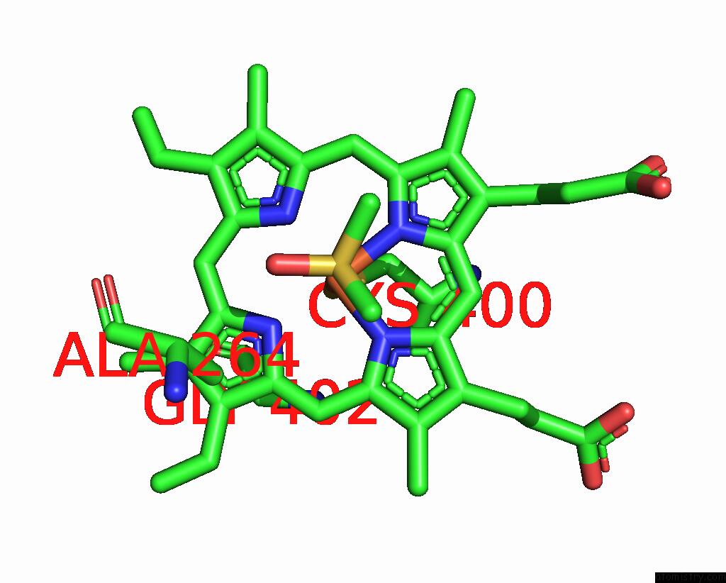
Mono view
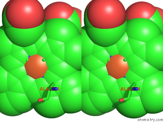
Stereo pair view
Mono view
Stereo pair view
A full contact list of Iron with other atoms in the Fe binding
site number 2 of Crystal Structure of P450BM3 Mutant with N-Perfluoroheptanoyl-L- Tryptophan within 5.0Å range:
|
Reference:
Z.Cong,
O.Shoji,
C.Kasai,
H.Sugimoto,
Y.Shiro,
Y.Watanabe.
Crystal Structure of P450BM3 with Decoy Molecules To Be Published.
Page generated: Tue Aug 5 19:10:39 2025
Last articles
Fe in 6MYQFe in 6MSO
Fe in 6MYP
Fe in 6MYO
Fe in 6MSN
Fe in 6MX5
Fe in 6MV0
Fe in 6MEV
Fe in 6MQ6
Fe in 6MQ1