Iron »
PDB 5b1a-5c1v »
5bv5 »
Iron in PDB 5bv5: Structure of CYP119 with T213A and C317H Mutations
Protein crystallography data
The structure of Structure of CYP119 with T213A and C317H Mutations, PDB code: 5bv5
was solved by
A.R.Buller,
T.Heel,
J.A.Mcintosh,
F.H.Arnold,
with X-Ray Crystallography technique. A brief refinement statistics is given in the table below:
| Resolution Low / High (Å) | 50.00 / 2.70 |
| Space group | P 1 21 1 |
| Cell size a, b, c (Å), α, β, γ (°) | 64.486, 137.871, 91.779, 90.00, 101.18, 90.00 |
| R / Rfree (%) | 23.8 / 27.7 |
Iron Binding Sites:
The binding sites of Iron atom in the Structure of CYP119 with T213A and C317H Mutations
(pdb code 5bv5). This binding sites where shown within
5.0 Angstroms radius around Iron atom.
In total 4 binding sites of Iron where determined in the Structure of CYP119 with T213A and C317H Mutations, PDB code: 5bv5:
Jump to Iron binding site number: 1; 2; 3; 4;
In total 4 binding sites of Iron where determined in the Structure of CYP119 with T213A and C317H Mutations, PDB code: 5bv5:
Jump to Iron binding site number: 1; 2; 3; 4;
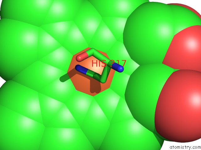
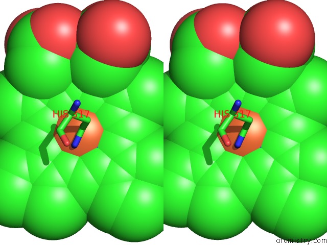
Iron binding site 1 out of 4 in 5bv5
Go back to
Iron binding site 1 out
of 4 in the Structure of CYP119 with T213A and C317H Mutations
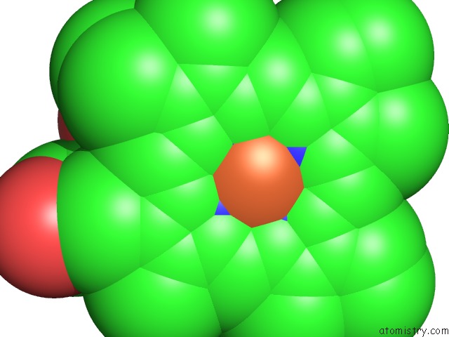
Mono view
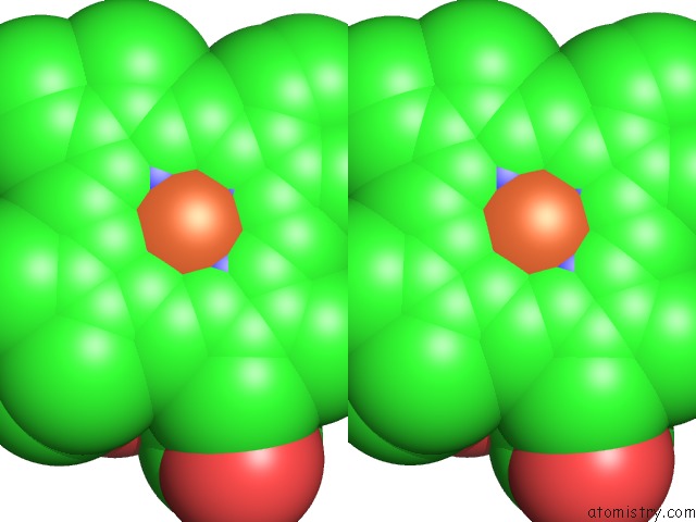
Stereo pair view
Mono view
Stereo pair view
A full contact list of Iron with other atoms in the Fe binding
site number 1 of Structure of CYP119 with T213A and C317H Mutations within 5.0Å range:
|
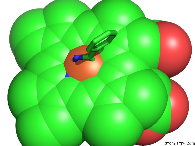
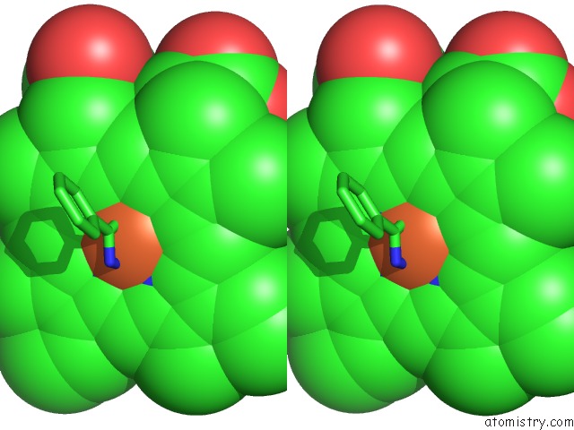
Iron binding site 2 out of 4 in 5bv5
Go back to
Iron binding site 2 out
of 4 in the Structure of CYP119 with T213A and C317H Mutations
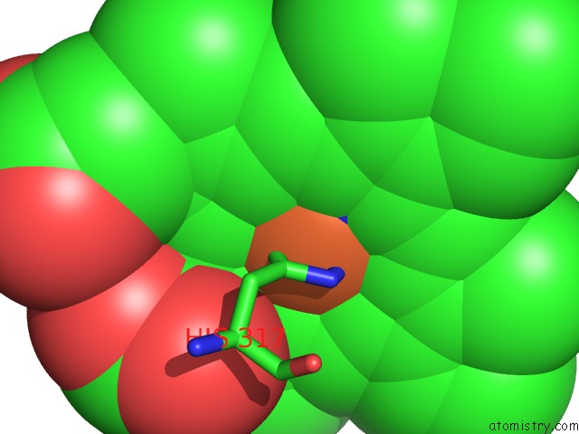
Mono view
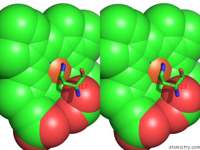
Stereo pair view
Mono view
Stereo pair view
A full contact list of Iron with other atoms in the Fe binding
site number 2 of Structure of CYP119 with T213A and C317H Mutations within 5.0Å range:
|
Iron binding site 3 out of 4 in 5bv5
Go back to
Iron binding site 3 out
of 4 in the Structure of CYP119 with T213A and C317H Mutations
Mono view
Stereo pair view
Mono view
Stereo pair view
A full contact list of Iron with other atoms in the Fe binding
site number 3 of Structure of CYP119 with T213A and C317H Mutations within 5.0Å range:
|
Iron binding site 4 out of 4 in 5bv5
Go back to
Iron binding site 4 out
of 4 in the Structure of CYP119 with T213A and C317H Mutations
Mono view
Stereo pair view
Mono view
Stereo pair view
A full contact list of Iron with other atoms in the Fe binding
site number 4 of Structure of CYP119 with T213A and C317H Mutations within 5.0Å range:
|
Reference:
J.A.Mcintosh,
T.Heel,
A.R.Buller,
L.Chio,
F.H.Arnold.
Structural Adaptability Facilitates Histidine Heme Ligation in A Cytochrome P450. J.Am.Chem.Soc. V. 137 13861 2015.
ISSN: ESSN 1520-5126
PubMed: 26299431
DOI: 10.1021/JACS.5B07107
Page generated: Mon Aug 5 20:08:15 2024
ISSN: ESSN 1520-5126
PubMed: 26299431
DOI: 10.1021/JACS.5B07107
Last articles
Zn in 9MJ5Zn in 9HNW
Zn in 9G0L
Zn in 9FNE
Zn in 9DZN
Zn in 9E0I
Zn in 9D32
Zn in 9DAK
Zn in 8ZXC
Zn in 8ZUF