Iron »
PDB 5c2i-5cnd »
5c3j »
Iron in PDB 5c3j: Crystal Structure of Mitochondrial Rhodoquinol-Fumarate Reductase From Ascaris Suum with Ubiquinone-1
Enzymatic activity of Crystal Structure of Mitochondrial Rhodoquinol-Fumarate Reductase From Ascaris Suum with Ubiquinone-1
All present enzymatic activity of Crystal Structure of Mitochondrial Rhodoquinol-Fumarate Reductase From Ascaris Suum with Ubiquinone-1:
1.3.5.1;
1.3.5.1;
Protein crystallography data
The structure of Crystal Structure of Mitochondrial Rhodoquinol-Fumarate Reductase From Ascaris Suum with Ubiquinone-1, PDB code: 5c3j
was solved by
S.Harada,
T.Shiba,
D.Sato,
A.Yamamoto,
M.Nagahama,
A.Yone,
D.K.Inaoka,
K.Sakamoto,
M.Inoue,
T.Honma,
K.Kita,
with X-Ray Crystallography technique. A brief refinement statistics is given in the table below:
| Resolution Low / High (Å) | 20.00 / 2.80 |
| Space group | P 21 21 21 |
| Cell size a, b, c (Å), α, β, γ (°) | 123.750, 126.827, 220.449, 90.00, 90.00, 90.00 |
| R / Rfree (%) | 20.2 / 26 |
Iron Binding Sites:
Pages:
>>> Page 1 <<< Page 2, Binding sites: 11 - 20;Binding sites:
The binding sites of Iron atom in the Crystal Structure of Mitochondrial Rhodoquinol-Fumarate Reductase From Ascaris Suum with Ubiquinone-1 (pdb code 5c3j). This binding sites where shown within 5.0 Angstroms radius around Iron atom.In total 20 binding sites of Iron where determined in the Crystal Structure of Mitochondrial Rhodoquinol-Fumarate Reductase From Ascaris Suum with Ubiquinone-1, PDB code: 5c3j:
Jump to Iron binding site number: 1; 2; 3; 4; 5; 6; 7; 8; 9; 10;
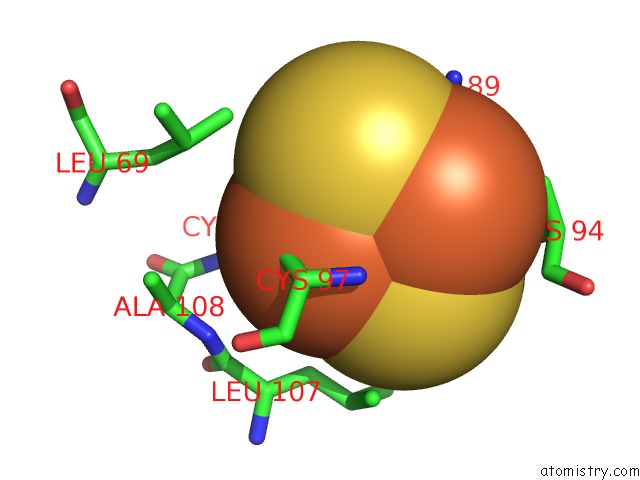
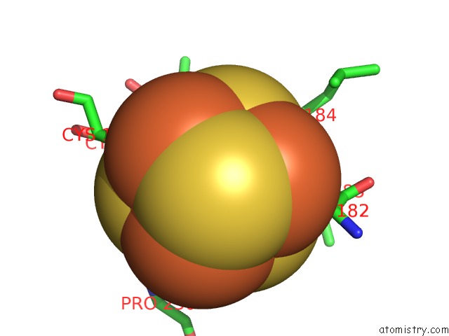
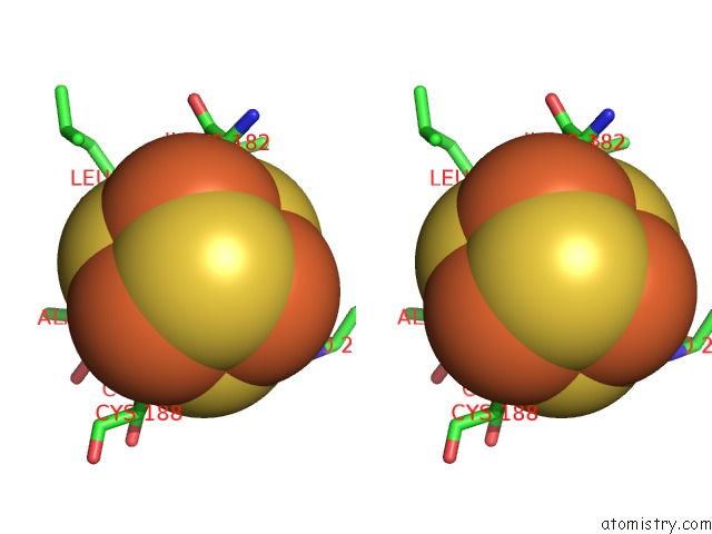
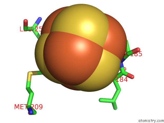
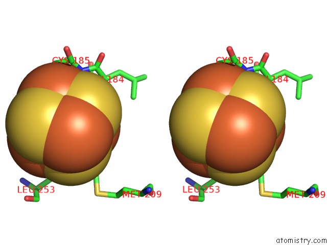
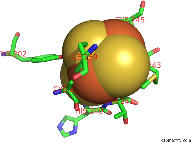
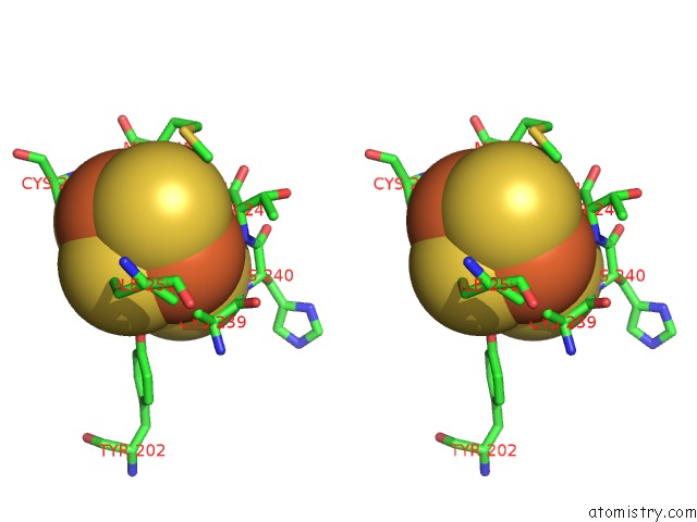
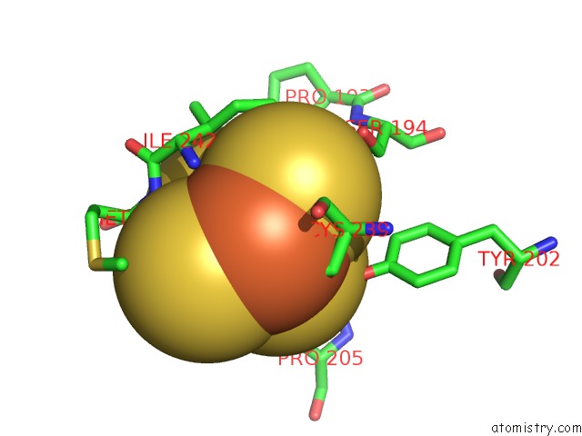
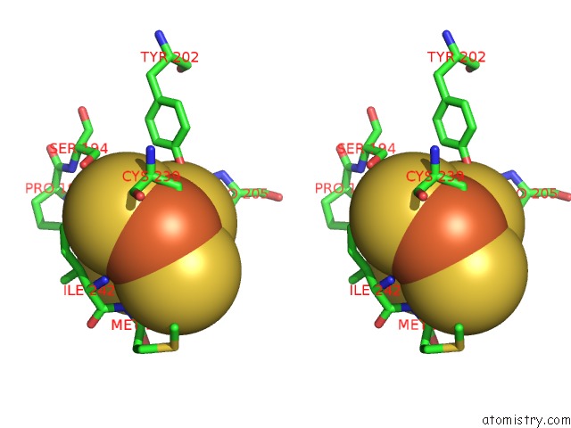
Iron binding site 1 out of 20 in 5c3j
Go back to
Iron binding site 1 out
of 20 in the Crystal Structure of Mitochondrial Rhodoquinol-Fumarate Reductase From Ascaris Suum with Ubiquinone-1
Mono view
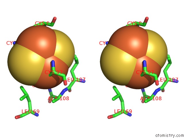
Stereo pair view
Mono view
Stereo pair view
A full contact list of Iron with other atoms in the Fe binding
site number 1 of Crystal Structure of Mitochondrial Rhodoquinol-Fumarate Reductase From Ascaris Suum with Ubiquinone-1 within 5.0Å range:
|
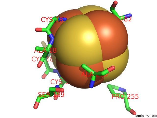
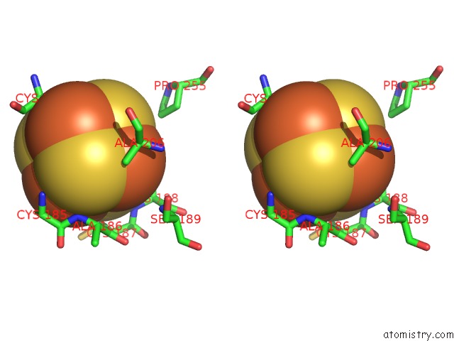
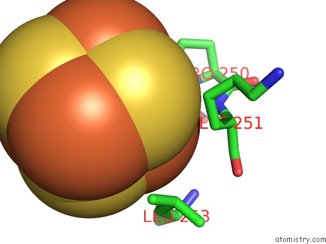
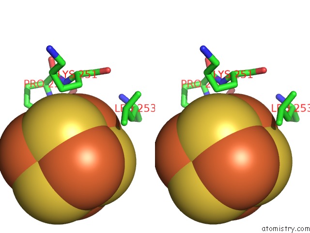
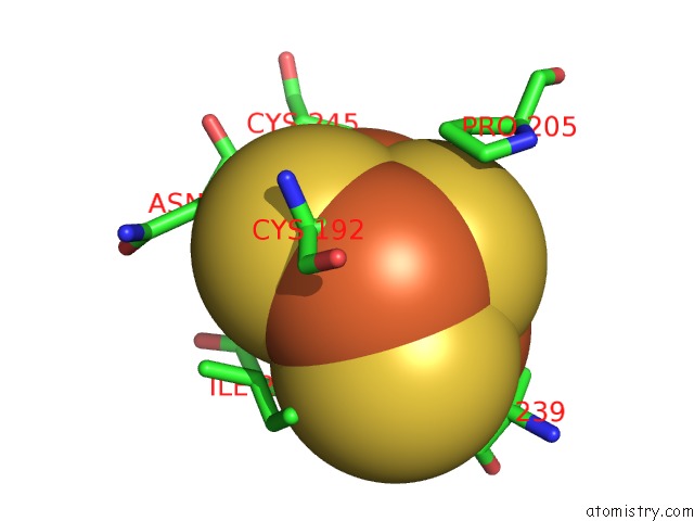
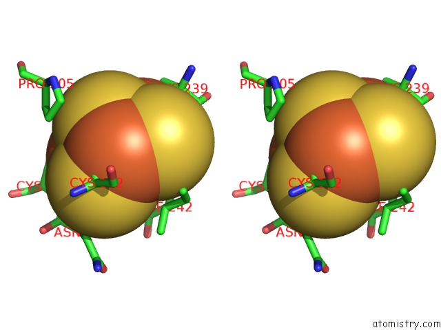
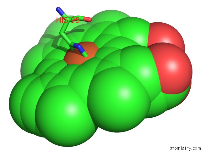
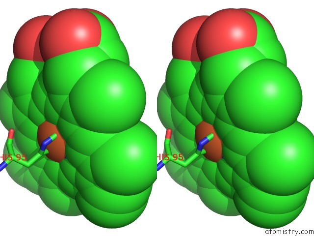
Iron binding site 2 out of 20 in 5c3j
Go back to
Iron binding site 2 out
of 20 in the Crystal Structure of Mitochondrial Rhodoquinol-Fumarate Reductase From Ascaris Suum with Ubiquinone-1
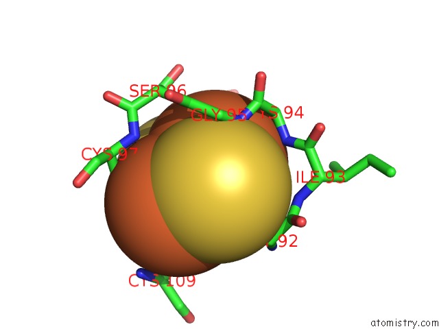
Mono view
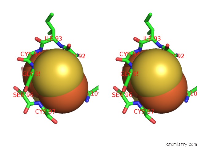
Stereo pair view
Mono view
Stereo pair view
A full contact list of Iron with other atoms in the Fe binding
site number 2 of Crystal Structure of Mitochondrial Rhodoquinol-Fumarate Reductase From Ascaris Suum with Ubiquinone-1 within 5.0Å range:
|
Iron binding site 3 out of 20 in 5c3j
Go back to
Iron binding site 3 out
of 20 in the Crystal Structure of Mitochondrial Rhodoquinol-Fumarate Reductase From Ascaris Suum with Ubiquinone-1
Mono view
Stereo pair view
Mono view
Stereo pair view
A full contact list of Iron with other atoms in the Fe binding
site number 3 of Crystal Structure of Mitochondrial Rhodoquinol-Fumarate Reductase From Ascaris Suum with Ubiquinone-1 within 5.0Å range:
|
Iron binding site 4 out of 20 in 5c3j
Go back to
Iron binding site 4 out
of 20 in the Crystal Structure of Mitochondrial Rhodoquinol-Fumarate Reductase From Ascaris Suum with Ubiquinone-1
Mono view
Stereo pair view
Mono view
Stereo pair view
A full contact list of Iron with other atoms in the Fe binding
site number 4 of Crystal Structure of Mitochondrial Rhodoquinol-Fumarate Reductase From Ascaris Suum with Ubiquinone-1 within 5.0Å range:
|
Iron binding site 5 out of 20 in 5c3j
Go back to
Iron binding site 5 out
of 20 in the Crystal Structure of Mitochondrial Rhodoquinol-Fumarate Reductase From Ascaris Suum with Ubiquinone-1
Mono view
Stereo pair view
Mono view
Stereo pair view
A full contact list of Iron with other atoms in the Fe binding
site number 5 of Crystal Structure of Mitochondrial Rhodoquinol-Fumarate Reductase From Ascaris Suum with Ubiquinone-1 within 5.0Å range:
|
Iron binding site 6 out of 20 in 5c3j
Go back to
Iron binding site 6 out
of 20 in the Crystal Structure of Mitochondrial Rhodoquinol-Fumarate Reductase From Ascaris Suum with Ubiquinone-1
Mono view
Stereo pair view
Mono view
Stereo pair view
A full contact list of Iron with other atoms in the Fe binding
site number 6 of Crystal Structure of Mitochondrial Rhodoquinol-Fumarate Reductase From Ascaris Suum with Ubiquinone-1 within 5.0Å range:
|
Iron binding site 7 out of 20 in 5c3j
Go back to
Iron binding site 7 out
of 20 in the Crystal Structure of Mitochondrial Rhodoquinol-Fumarate Reductase From Ascaris Suum with Ubiquinone-1
Mono view
Stereo pair view
Mono view
Stereo pair view
A full contact list of Iron with other atoms in the Fe binding
site number 7 of Crystal Structure of Mitochondrial Rhodoquinol-Fumarate Reductase From Ascaris Suum with Ubiquinone-1 within 5.0Å range:
|
Iron binding site 8 out of 20 in 5c3j
Go back to
Iron binding site 8 out
of 20 in the Crystal Structure of Mitochondrial Rhodoquinol-Fumarate Reductase From Ascaris Suum with Ubiquinone-1
Mono view
Stereo pair view
Mono view
Stereo pair view
A full contact list of Iron with other atoms in the Fe binding
site number 8 of Crystal Structure of Mitochondrial Rhodoquinol-Fumarate Reductase From Ascaris Suum with Ubiquinone-1 within 5.0Å range:
|
Iron binding site 9 out of 20 in 5c3j
Go back to
Iron binding site 9 out
of 20 in the Crystal Structure of Mitochondrial Rhodoquinol-Fumarate Reductase From Ascaris Suum with Ubiquinone-1
Mono view
Stereo pair view
Mono view
Stereo pair view
A full contact list of Iron with other atoms in the Fe binding
site number 9 of Crystal Structure of Mitochondrial Rhodoquinol-Fumarate Reductase From Ascaris Suum with Ubiquinone-1 within 5.0Å range:
|
Iron binding site 10 out of 20 in 5c3j
Go back to
Iron binding site 10 out
of 20 in the Crystal Structure of Mitochondrial Rhodoquinol-Fumarate Reductase From Ascaris Suum with Ubiquinone-1
Mono view
Stereo pair view
Mono view
Stereo pair view
A full contact list of Iron with other atoms in the Fe binding
site number 10 of Crystal Structure of Mitochondrial Rhodoquinol-Fumarate Reductase From Ascaris Suum with Ubiquinone-1 within 5.0Å range:
|
Reference:
S.Harada,
T.Shiba,
D.Sato,
A.Yamamoto,
M.Nagahama,
A.Yone,
D.K.Inaoka,
K.Sakamoto,
M.Inoue,
T.Honma,
K.Kita.
Structural Insights Into the Molecular Design of Flutolanil Derivatives Targeted For Fumarate Respiration of Parasite Mitochondria To Be Published.
Page generated: Mon Aug 5 20:48:05 2024
Last articles
Zn in 9JYWZn in 9IR4
Zn in 9IR3
Zn in 9GMX
Zn in 9GMW
Zn in 9JEJ
Zn in 9ERF
Zn in 9ERE
Zn in 9EGV
Zn in 9EGW