Iron »
PDB 5c2i-5cnd »
5c6f »
Iron in PDB 5c6f: Crystal Structures of Ferritin Mutants Reveal Side-on Binding to Diiron and End-on Cleavage of Oxygen
Enzymatic activity of Crystal Structures of Ferritin Mutants Reveal Side-on Binding to Diiron and End-on Cleavage of Oxygen
All present enzymatic activity of Crystal Structures of Ferritin Mutants Reveal Side-on Binding to Diiron and End-on Cleavage of Oxygen:
1.16.3.2;
1.16.3.2;
Protein crystallography data
The structure of Crystal Structures of Ferritin Mutants Reveal Side-on Binding to Diiron and End-on Cleavage of Oxygen, PDB code: 5c6f
was solved by
S.Kim,
K.H.Kim,
J.H.Seok,
Y.H.Park,
S.W.Jung,
Y.B.Chung,
D.B.Lee,
J.H.Lee,
K.R.Han,
A.E.Cho,
C.Lee,
M.S.Chung,
with X-Ray Crystallography technique. A brief refinement statistics is given in the table below:
| Resolution Low / High (Å) | 50.00 / 2.00 |
| Space group | P 4 |
| Cell size a, b, c (Å), α, β, γ (°) | 128.051, 128.051, 165.103, 90.00, 90.00, 90.00 |
| R / Rfree (%) | 14.9 / 19.5 |
Iron Binding Sites:
The binding sites of Iron atom in the Crystal Structures of Ferritin Mutants Reveal Side-on Binding to Diiron and End-on Cleavage of Oxygen
(pdb code 5c6f). This binding sites where shown within
5.0 Angstroms radius around Iron atom.
In total 9 binding sites of Iron where determined in the Crystal Structures of Ferritin Mutants Reveal Side-on Binding to Diiron and End-on Cleavage of Oxygen, PDB code: 5c6f:
Jump to Iron binding site number: 1; 2; 3; 4; 5; 6; 7; 8; 9;
In total 9 binding sites of Iron where determined in the Crystal Structures of Ferritin Mutants Reveal Side-on Binding to Diiron and End-on Cleavage of Oxygen, PDB code: 5c6f:
Jump to Iron binding site number: 1; 2; 3; 4; 5; 6; 7; 8; 9;
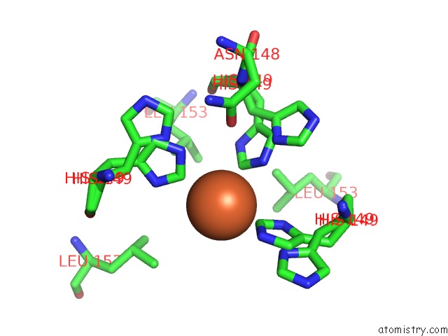
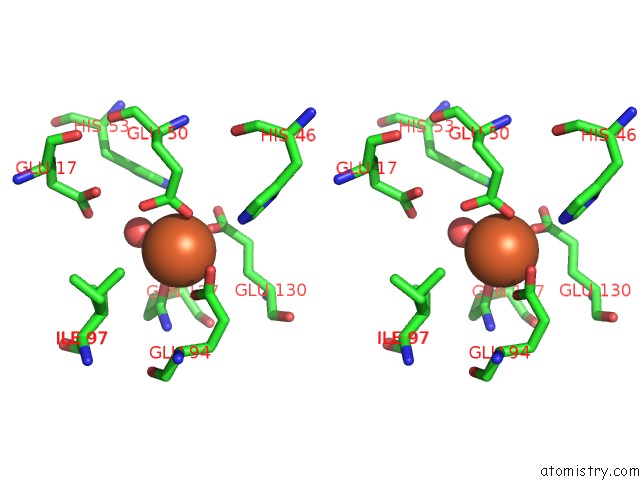
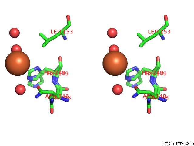
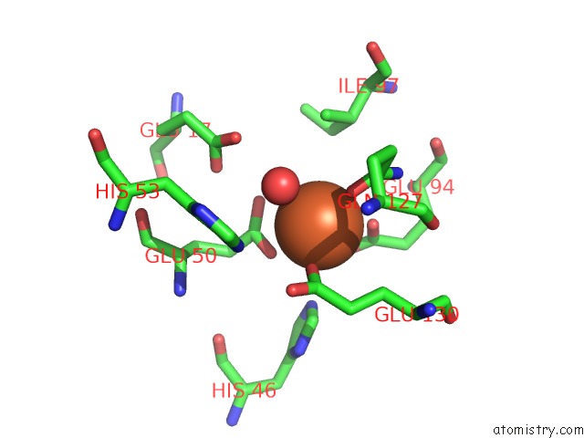
Iron binding site 1 out of 9 in 5c6f
Go back to
Iron binding site 1 out
of 9 in the Crystal Structures of Ferritin Mutants Reveal Side-on Binding to Diiron and End-on Cleavage of Oxygen
Mono view
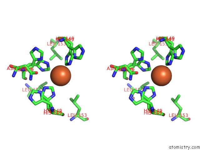
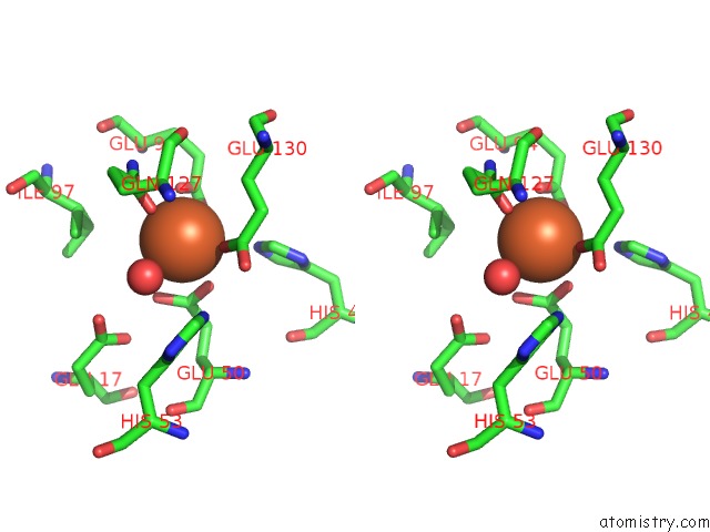
Stereo pair view
Mono view
Stereo pair view
A full contact list of Iron with other atoms in the Fe binding
site number 1 of Crystal Structures of Ferritin Mutants Reveal Side-on Binding to Diiron and End-on Cleavage of Oxygen within 5.0Å range:
|
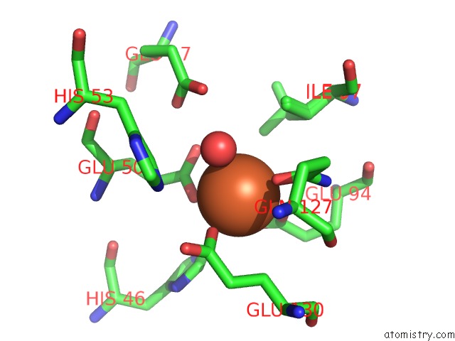
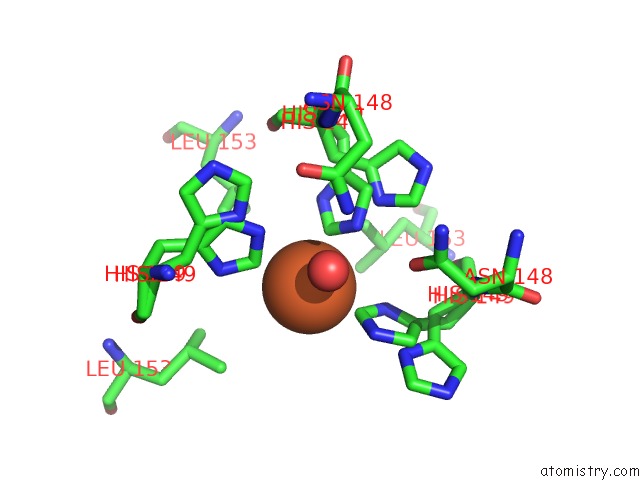
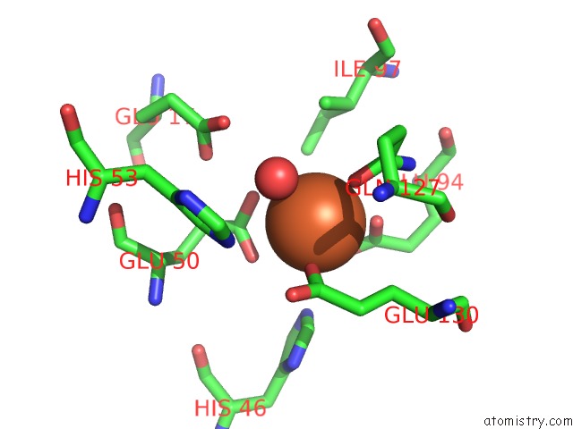
Iron binding site 2 out of 9 in 5c6f
Go back to
Iron binding site 2 out
of 9 in the Crystal Structures of Ferritin Mutants Reveal Side-on Binding to Diiron and End-on Cleavage of Oxygen

Mono view
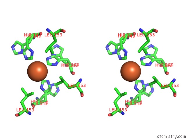
Stereo pair view
Mono view
Stereo pair view
A full contact list of Iron with other atoms in the Fe binding
site number 2 of Crystal Structures of Ferritin Mutants Reveal Side-on Binding to Diiron and End-on Cleavage of Oxygen within 5.0Å range:
|
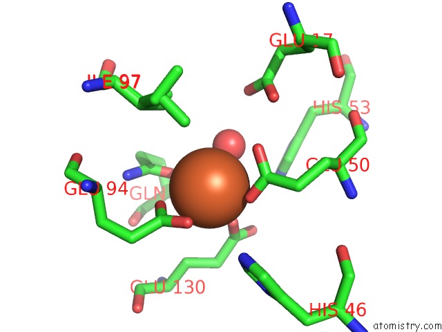
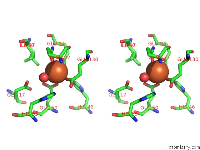
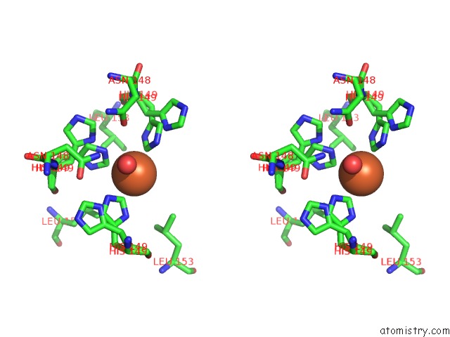
Iron binding site 3 out of 9 in 5c6f
Go back to
Iron binding site 3 out
of 9 in the Crystal Structures of Ferritin Mutants Reveal Side-on Binding to Diiron and End-on Cleavage of Oxygen
Mono view
Stereo pair view
Mono view
Stereo pair view
A full contact list of Iron with other atoms in the Fe binding
site number 3 of Crystal Structures of Ferritin Mutants Reveal Side-on Binding to Diiron and End-on Cleavage of Oxygen within 5.0Å range:
|
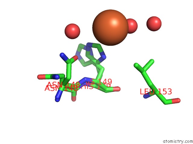
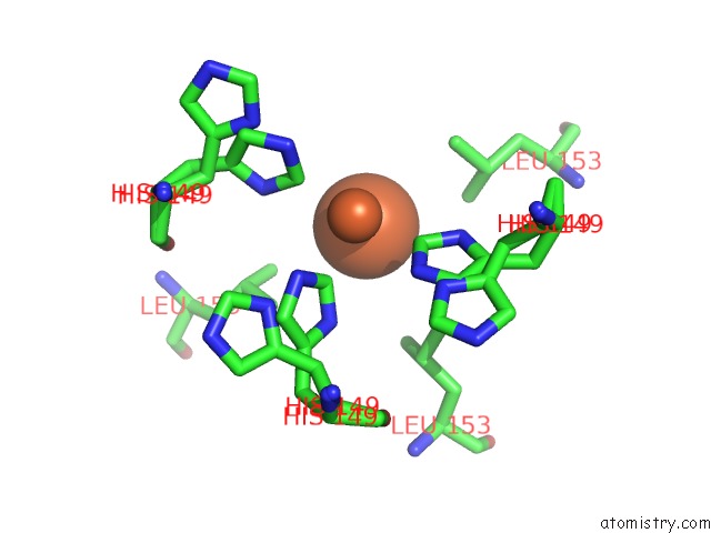
Iron binding site 4 out of 9 in 5c6f
Go back to
Iron binding site 4 out
of 9 in the Crystal Structures of Ferritin Mutants Reveal Side-on Binding to Diiron and End-on Cleavage of Oxygen
Mono view
Stereo pair view
Mono view
Stereo pair view
A full contact list of Iron with other atoms in the Fe binding
site number 4 of Crystal Structures of Ferritin Mutants Reveal Side-on Binding to Diiron and End-on Cleavage of Oxygen within 5.0Å range:
|
Iron binding site 5 out of 9 in 5c6f
Go back to
Iron binding site 5 out
of 9 in the Crystal Structures of Ferritin Mutants Reveal Side-on Binding to Diiron and End-on Cleavage of Oxygen
Mono view
Stereo pair view
Mono view
Stereo pair view
A full contact list of Iron with other atoms in the Fe binding
site number 5 of Crystal Structures of Ferritin Mutants Reveal Side-on Binding to Diiron and End-on Cleavage of Oxygen within 5.0Å range:
|
Iron binding site 6 out of 9 in 5c6f
Go back to
Iron binding site 6 out
of 9 in the Crystal Structures of Ferritin Mutants Reveal Side-on Binding to Diiron and End-on Cleavage of Oxygen
Mono view
Stereo pair view
Mono view
Stereo pair view
A full contact list of Iron with other atoms in the Fe binding
site number 6 of Crystal Structures of Ferritin Mutants Reveal Side-on Binding to Diiron and End-on Cleavage of Oxygen within 5.0Å range:
|
Iron binding site 7 out of 9 in 5c6f
Go back to
Iron binding site 7 out
of 9 in the Crystal Structures of Ferritin Mutants Reveal Side-on Binding to Diiron and End-on Cleavage of Oxygen
Mono view

Stereo pair view
Mono view
Stereo pair view
A full contact list of Iron with other atoms in the Fe binding
site number 7 of Crystal Structures of Ferritin Mutants Reveal Side-on Binding to Diiron and End-on Cleavage of Oxygen within 5.0Å range:
|
Iron binding site 8 out of 9 in 5c6f
Go back to
Iron binding site 8 out
of 9 in the Crystal Structures of Ferritin Mutants Reveal Side-on Binding to Diiron and End-on Cleavage of Oxygen
Mono view
Stereo pair view
Mono view
Stereo pair view
A full contact list of Iron with other atoms in the Fe binding
site number 8 of Crystal Structures of Ferritin Mutants Reveal Side-on Binding to Diiron and End-on Cleavage of Oxygen within 5.0Å range:
|
Iron binding site 9 out of 9 in 5c6f
Go back to
Iron binding site 9 out
of 9 in the Crystal Structures of Ferritin Mutants Reveal Side-on Binding to Diiron and End-on Cleavage of Oxygen
Mono view

Stereo pair view
Mono view
Stereo pair view
A full contact list of Iron with other atoms in the Fe binding
site number 9 of Crystal Structures of Ferritin Mutants Reveal Side-on Binding to Diiron and End-on Cleavage of Oxygen within 5.0Å range:
|
Reference:
S.Kim,
J.H.Lee,
J.H.Seok,
Y.H.Park,
S.W.Jung,
A.E.Cho,
C.Lee,
M.S.Chung,
K.H.Kim.
Structural Basis of Novel Iron-Uptake Route and Reaction Intermediates in Ferritins From Gram-Negative Bacteria. J. Mol. Biol. V. 428 5007 2016.
ISSN: ESSN 1089-8638
PubMed: 27777002
DOI: 10.1016/J.JMB.2016.10.022
Page generated: Mon Aug 5 20:48:05 2024
ISSN: ESSN 1089-8638
PubMed: 27777002
DOI: 10.1016/J.JMB.2016.10.022
Last articles
F in 7M8VF in 7MCK
F in 7MAZ
F in 7MBO
F in 7MCE
F in 7MB2
F in 7MB1
F in 7MAX
F in 7M8R
F in 7M9R