Iron »
PDB 5c2i-5cnd »
5cjh »
Iron in PDB 5cjh: Crystal Structure of Eukaryotic Oxoiron MAGKATG2 at pH 8.5
Enzymatic activity of Crystal Structure of Eukaryotic Oxoiron MAGKATG2 at pH 8.5
All present enzymatic activity of Crystal Structure of Eukaryotic Oxoiron MAGKATG2 at pH 8.5:
1.11.1.21;
1.11.1.21;
Protein crystallography data
The structure of Crystal Structure of Eukaryotic Oxoiron MAGKATG2 at pH 8.5, PDB code: 5cjh
was solved by
B.Gasselhuber,
C.Obinger,
I.Fita,
X.Carpena,
with X-Ray Crystallography technique. A brief refinement statistics is given in the table below:
| Resolution Low / High (Å) | 19.94 / 1.60 |
| Space group | P 21 21 21 |
| Cell size a, b, c (Å), α, β, γ (°) | 103.530, 109.540, 132.150, 90.00, 90.00, 90.00 |
| R / Rfree (%) | 16.5 / 19 |
Iron Binding Sites:
The binding sites of Iron atom in the Crystal Structure of Eukaryotic Oxoiron MAGKATG2 at pH 8.5
(pdb code 5cjh). This binding sites where shown within
5.0 Angstroms radius around Iron atom.
In total 2 binding sites of Iron where determined in the Crystal Structure of Eukaryotic Oxoiron MAGKATG2 at pH 8.5, PDB code: 5cjh:
Jump to Iron binding site number: 1; 2;
In total 2 binding sites of Iron where determined in the Crystal Structure of Eukaryotic Oxoiron MAGKATG2 at pH 8.5, PDB code: 5cjh:
Jump to Iron binding site number: 1; 2;
Iron binding site 1 out of 2 in 5cjh
Go back to
Iron binding site 1 out
of 2 in the Crystal Structure of Eukaryotic Oxoiron MAGKATG2 at pH 8.5
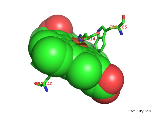
Mono view
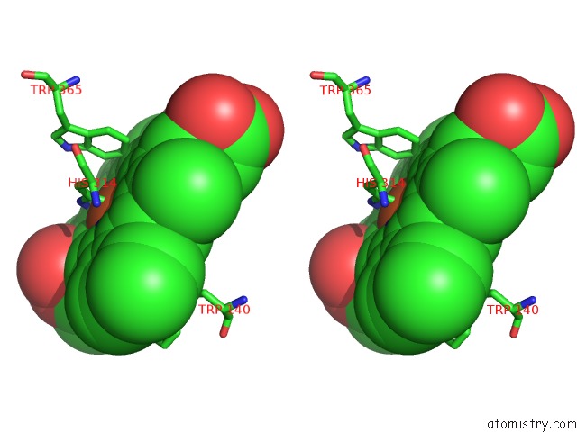
Stereo pair view
Mono view
Stereo pair view
A full contact list of Iron with other atoms in the Fe binding
site number 1 of Crystal Structure of Eukaryotic Oxoiron MAGKATG2 at pH 8.5 within 5.0Å range:
|
Iron binding site 2 out of 2 in 5cjh
Go back to
Iron binding site 2 out
of 2 in the Crystal Structure of Eukaryotic Oxoiron MAGKATG2 at pH 8.5
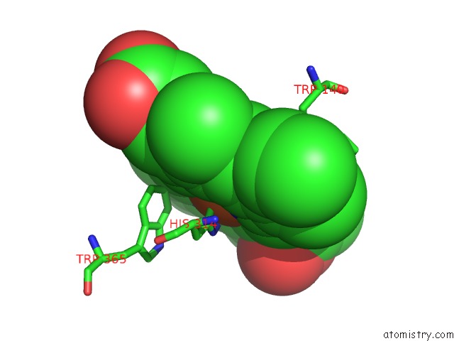
Mono view
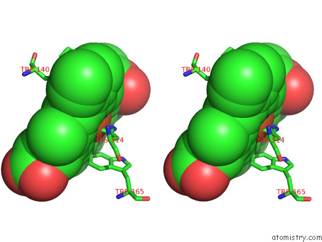
Stereo pair view
Mono view
Stereo pair view
A full contact list of Iron with other atoms in the Fe binding
site number 2 of Crystal Structure of Eukaryotic Oxoiron MAGKATG2 at pH 8.5 within 5.0Å range:
|
Reference:
B.Gasselhuber,
X.Carpena,
M.M.Graf,
K.F.Pirker,
A.Nicolussi,
A.Sundermann,
S.Hofbauer,
M.Zamocky,
P.G.Furtmuller,
C.Jakopitsch,
C.Oostenbrink,
I.Fita,
C.Obinger.
Eukaryotic Catalase-Peroxidase: the Role of the Trp-Tyr-Met Adduct in Protein Stability, Substrate Accessibility, and Catalysis of Hydrogen Peroxide Dismutation. Biochemistry V. 54 5425 2015.
ISSN: ISSN 0006-2960
PubMed: 26290940
DOI: 10.1021/ACS.BIOCHEM.5B00831
Page generated: Mon Aug 5 21:06:46 2024
ISSN: ISSN 0006-2960
PubMed: 26290940
DOI: 10.1021/ACS.BIOCHEM.5B00831
Last articles
Fe in 2YXOFe in 2YRS
Fe in 2YXC
Fe in 2YNM
Fe in 2YVJ
Fe in 2YP1
Fe in 2YU2
Fe in 2YU1
Fe in 2YQB
Fe in 2YOO