Iron »
PDB 5cne-5da5 »
5cvr »
Iron in PDB 5cvr: Crystal Structure of Fnr of A. Fischeri in A Partially Degraded Form
Protein crystallography data
The structure of Crystal Structure of Fnr of A. Fischeri in A Partially Degraded Form, PDB code: 5cvr
was solved by
A.Volbeda,
J.C.Fontecilla-Camps,
with X-Ray Crystallography technique. A brief refinement statistics is given in the table below:
| Resolution Low / High (Å) | 37.64 / 2.60 |
| Space group | I 4 2 2 |
| Cell size a, b, c (Å), α, β, γ (°) | 75.270, 75.270, 212.430, 90.00, 90.00, 90.00 |
| R / Rfree (%) | 19.1 / 23.7 |
Iron Binding Sites:
The binding sites of Iron atom in the Crystal Structure of Fnr of A. Fischeri in A Partially Degraded Form
(pdb code 5cvr). This binding sites where shown within
5.0 Angstroms radius around Iron atom.
In total 2 binding sites of Iron where determined in the Crystal Structure of Fnr of A. Fischeri in A Partially Degraded Form, PDB code: 5cvr:
Jump to Iron binding site number: 1; 2;
In total 2 binding sites of Iron where determined in the Crystal Structure of Fnr of A. Fischeri in A Partially Degraded Form, PDB code: 5cvr:
Jump to Iron binding site number: 1; 2;
Iron binding site 1 out of 2 in 5cvr
Go back to
Iron binding site 1 out
of 2 in the Crystal Structure of Fnr of A. Fischeri in A Partially Degraded Form
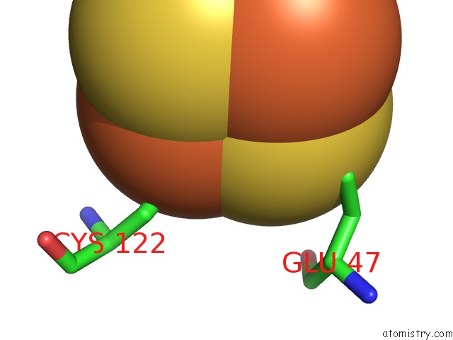
Mono view
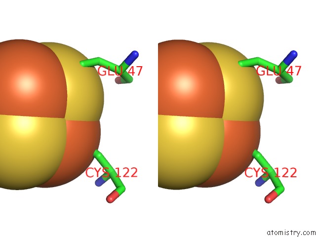
Stereo pair view
Mono view
Stereo pair view
A full contact list of Iron with other atoms in the Fe binding
site number 1 of Crystal Structure of Fnr of A. Fischeri in A Partially Degraded Form within 5.0Å range:
|
Iron binding site 2 out of 2 in 5cvr
Go back to
Iron binding site 2 out
of 2 in the Crystal Structure of Fnr of A. Fischeri in A Partially Degraded Form
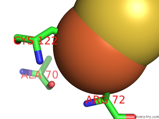
Mono view
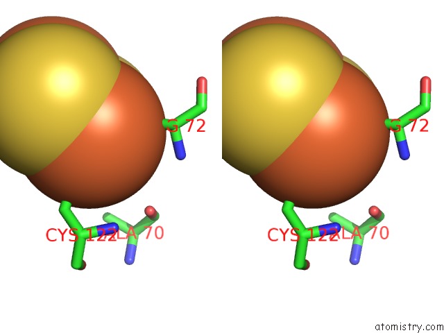
Stereo pair view
Mono view
Stereo pair view
A full contact list of Iron with other atoms in the Fe binding
site number 2 of Crystal Structure of Fnr of A. Fischeri in A Partially Degraded Form within 5.0Å range:
|
Reference:
A.Volbeda,
C.Darnault,
O.Renoux,
Y.Nicolet,
J.C.Fontecilla-Camps.
The Crystal Structure of the Global Anaerobic Transcriptional Regulator Fnr Explains Its Extremely Fine-Tuned Monomer-Dimer Equilibrium. Sci Adv V. 1 01086 2015.
ISSN: ESSN 2375-2548
PubMed: 26665177
DOI: 10.1126/SCIADV.1501086
Page generated: Mon Aug 5 21:35:39 2024
ISSN: ESSN 2375-2548
PubMed: 26665177
DOI: 10.1126/SCIADV.1501086
Last articles
Zn in 9J0NZn in 9J0O
Zn in 9J0P
Zn in 9FJX
Zn in 9EKB
Zn in 9C0F
Zn in 9CAH
Zn in 9CH0
Zn in 9CH3
Zn in 9CH1