Iron »
PDB 5dab-5eax »
5dco »
Iron in PDB 5dco: R2-Like Ligand-Binding Oxidase with Aerobically Reconstituted Diiron Cofactor (Short Soak)
Enzymatic activity of R2-Like Ligand-Binding Oxidase with Aerobically Reconstituted Diiron Cofactor (Short Soak)
All present enzymatic activity of R2-Like Ligand-Binding Oxidase with Aerobically Reconstituted Diiron Cofactor (Short Soak):
1.17.4.1;
1.17.4.1;
Protein crystallography data
The structure of R2-Like Ligand-Binding Oxidase with Aerobically Reconstituted Diiron Cofactor (Short Soak), PDB code: 5dco
was solved by
J.J.Griese,
M.Hogbom,
with X-Ray Crystallography technique. A brief refinement statistics is given in the table below:
| Resolution Low / High (Å) | 48.22 / 2.33 |
| Space group | I 2 2 2 |
| Cell size a, b, c (Å), α, β, γ (°) | 55.629, 96.695, 128.342, 90.00, 90.00, 90.00 |
| R / Rfree (%) | 15.7 / 22.6 |
Iron Binding Sites:
The binding sites of Iron atom in the R2-Like Ligand-Binding Oxidase with Aerobically Reconstituted Diiron Cofactor (Short Soak)
(pdb code 5dco). This binding sites where shown within
5.0 Angstroms radius around Iron atom.
In total 2 binding sites of Iron where determined in the R2-Like Ligand-Binding Oxidase with Aerobically Reconstituted Diiron Cofactor (Short Soak), PDB code: 5dco:
Jump to Iron binding site number: 1; 2;
In total 2 binding sites of Iron where determined in the R2-Like Ligand-Binding Oxidase with Aerobically Reconstituted Diiron Cofactor (Short Soak), PDB code: 5dco:
Jump to Iron binding site number: 1; 2;
Iron binding site 1 out of 2 in 5dco
Go back to
Iron binding site 1 out
of 2 in the R2-Like Ligand-Binding Oxidase with Aerobically Reconstituted Diiron Cofactor (Short Soak)
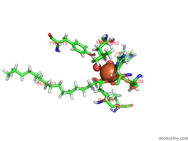
Mono view
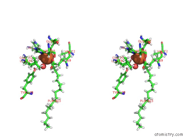
Stereo pair view
Mono view
Stereo pair view
A full contact list of Iron with other atoms in the Fe binding
site number 1 of R2-Like Ligand-Binding Oxidase with Aerobically Reconstituted Diiron Cofactor (Short Soak) within 5.0Å range:
|
Iron binding site 2 out of 2 in 5dco
Go back to
Iron binding site 2 out
of 2 in the R2-Like Ligand-Binding Oxidase with Aerobically Reconstituted Diiron Cofactor (Short Soak)
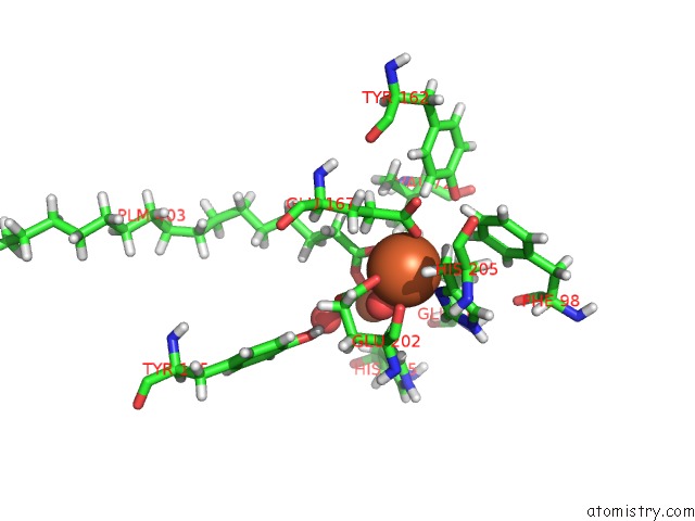
Mono view
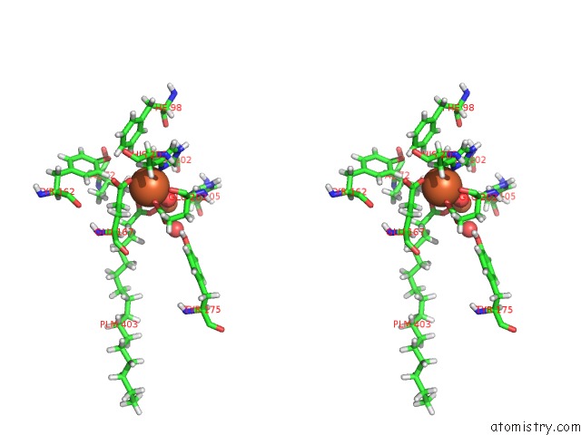
Stereo pair view
Mono view
Stereo pair view
A full contact list of Iron with other atoms in the Fe binding
site number 2 of R2-Like Ligand-Binding Oxidase with Aerobically Reconstituted Diiron Cofactor (Short Soak) within 5.0Å range:
|
Reference:
J.J.Griese,
R.Kositzki,
P.Schrapers,
R.M.Branca,
A.Nordstrom,
J.Lehtio,
M.Haumann,
M.Hogbom.
Structural Basis For Oxygen Activation at A Heterodinuclear Manganese/Iron Cofactor. J.Biol.Chem. V. 290 25254 2015.
ISSN: ESSN 1083-351X
PubMed: 26324712
DOI: 10.1074/JBC.M115.675223
Page generated: Mon Aug 5 23:18:17 2024
ISSN: ESSN 1083-351X
PubMed: 26324712
DOI: 10.1074/JBC.M115.675223
Last articles
Fe in 2YXOFe in 2YRS
Fe in 2YXC
Fe in 2YNM
Fe in 2YVJ
Fe in 2YP1
Fe in 2YU2
Fe in 2YU1
Fe in 2YQB
Fe in 2YOO