Iron »
PDB 5edt-5ex9 »
5ekb »
Iron in PDB 5ekb: R2-Like Ligand-Binding Oxidase with Aerobically Reconstituted Mn/Fe Cofactor (Reconstituted in Solution)
Enzymatic activity of R2-Like Ligand-Binding Oxidase with Aerobically Reconstituted Mn/Fe Cofactor (Reconstituted in Solution)
All present enzymatic activity of R2-Like Ligand-Binding Oxidase with Aerobically Reconstituted Mn/Fe Cofactor (Reconstituted in Solution):
1.17.4.1;
1.17.4.1;
Protein crystallography data
The structure of R2-Like Ligand-Binding Oxidase with Aerobically Reconstituted Mn/Fe Cofactor (Reconstituted in Solution), PDB code: 5ekb
was solved by
J.J.Griese,
M.Hogbom,
with X-Ray Crystallography technique. A brief refinement statistics is given in the table below:
| Resolution Low / High (Å) | 48.61 / 2.07 |
| Space group | I 2 2 2 |
| Cell size a, b, c (Å), α, β, γ (°) | 56.041, 97.217, 128.279, 90.00, 90.00, 90.00 |
| R / Rfree (%) | 19 / 24.4 |
Other elements in 5ekb:
The structure of R2-Like Ligand-Binding Oxidase with Aerobically Reconstituted Mn/Fe Cofactor (Reconstituted in Solution) also contains other interesting chemical elements:
| Manganese | (Mn) | 1 atom |
Iron Binding Sites:
The binding sites of Iron atom in the R2-Like Ligand-Binding Oxidase with Aerobically Reconstituted Mn/Fe Cofactor (Reconstituted in Solution)
(pdb code 5ekb). This binding sites where shown within
5.0 Angstroms radius around Iron atom.
In total only one binding site of Iron was determined in the R2-Like Ligand-Binding Oxidase with Aerobically Reconstituted Mn/Fe Cofactor (Reconstituted in Solution), PDB code: 5ekb:
In total only one binding site of Iron was determined in the R2-Like Ligand-Binding Oxidase with Aerobically Reconstituted Mn/Fe Cofactor (Reconstituted in Solution), PDB code: 5ekb:
Iron binding site 1 out of 1 in 5ekb
Go back to
Iron binding site 1 out
of 1 in the R2-Like Ligand-Binding Oxidase with Aerobically Reconstituted Mn/Fe Cofactor (Reconstituted in Solution)
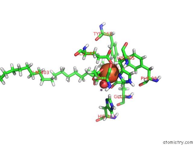
Mono view
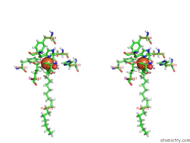
Stereo pair view
Mono view
Stereo pair view
A full contact list of Iron with other atoms in the Fe binding
site number 1 of R2-Like Ligand-Binding Oxidase with Aerobically Reconstituted Mn/Fe Cofactor (Reconstituted in Solution) within 5.0Å range:
|
Reference:
Y.Kutin,
V.Srinivas,
M.Fritz,
R.Kositzki,
H.S.Shafaat,
J.Birrell,
E.Bill,
M.Haumann,
W.Lubitz,
M.Hogbom,
J.J.Griese,
N.Cox.
Divergent Assembly Mechanisms of the Manganese/Iron Cofactors in R2LOX and R2C Proteins. J.Inorg.Biochem. V. 162 164 2016.
ISSN: ISSN 0162-0134
PubMed: 27138102
DOI: 10.1016/J.JINORGBIO.2016.04.019
Page generated: Tue Aug 6 00:29:04 2024
ISSN: ISSN 0162-0134
PubMed: 27138102
DOI: 10.1016/J.JINORGBIO.2016.04.019
Last articles
Zn in 9MJ5Zn in 9HNW
Zn in 9G0L
Zn in 9FNE
Zn in 9DZN
Zn in 9E0I
Zn in 9D32
Zn in 9DAK
Zn in 8ZXC
Zn in 8ZUF