Iron »
PDB 5edt-5ex9 »
5eui »
Iron in PDB 5eui: Structure of Predicted Ancestral Pika Hemoglobin
Protein crystallography data
The structure of Structure of Predicted Ancestral Pika Hemoglobin, PDB code: 5eui
was solved by
N.Inoguchi,
N.Chandrasekhar,
J.F.Storz,
H.Moriyama,
with X-Ray Crystallography technique. A brief refinement statistics is given in the table below:
| Resolution Low / High (Å) | 30.29 / 1.45 |
| Space group | C 2 2 21 |
| Cell size a, b, c (Å), α, β, γ (°) | 63.350, 91.412, 103.537, 90.00, 90.00, 90.00 |
| R / Rfree (%) | 19.4 / 21.1 |
Iron Binding Sites:
The binding sites of Iron atom in the Structure of Predicted Ancestral Pika Hemoglobin
(pdb code 5eui). This binding sites where shown within
5.0 Angstroms radius around Iron atom.
In total 2 binding sites of Iron where determined in the Structure of Predicted Ancestral Pika Hemoglobin, PDB code: 5eui:
Jump to Iron binding site number: 1; 2;
In total 2 binding sites of Iron where determined in the Structure of Predicted Ancestral Pika Hemoglobin, PDB code: 5eui:
Jump to Iron binding site number: 1; 2;
Iron binding site 1 out of 2 in 5eui
Go back to
Iron binding site 1 out
of 2 in the Structure of Predicted Ancestral Pika Hemoglobin
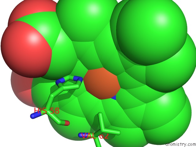
Mono view
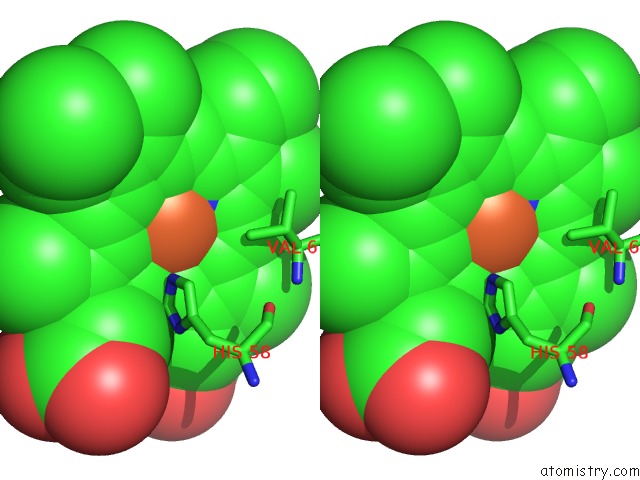
Stereo pair view
Mono view
Stereo pair view
A full contact list of Iron with other atoms in the Fe binding
site number 1 of Structure of Predicted Ancestral Pika Hemoglobin within 5.0Å range:
|
Iron binding site 2 out of 2 in 5eui
Go back to
Iron binding site 2 out
of 2 in the Structure of Predicted Ancestral Pika Hemoglobin
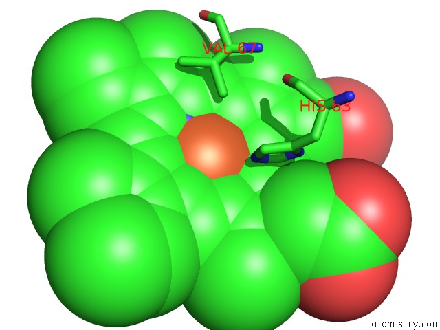
Mono view
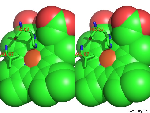
Stereo pair view
Mono view
Stereo pair view
A full contact list of Iron with other atoms in the Fe binding
site number 2 of Structure of Predicted Ancestral Pika Hemoglobin within 5.0Å range:
|
Reference:
N.Inoguchi,
C.Natarajan,
J.F.Storz,
H.Moriyama.
Structure of Predicted Pika Ancestral Hemoglobin at 1.45 Angstroms Resolution. To Be Published.
Page generated: Tue Aug 5 21:12:16 2025
Last articles
Fe in 5MDXFe in 5MCS
Fe in 5MAA
Fe in 5MAU
Fe in 5MAB
Fe in 5MBN
Fe in 5MBA
Fe in 5MAP
Fe in 5MA2
Fe in 5MA1