Iron »
PDB 5exd-5foi »
5exk »
Iron in PDB 5exk: Crystal Structure of M. Tuberculosis Lipoyl Synthase with 6- Thiooctanoyl Peptide Intermediate
Enzymatic activity of Crystal Structure of M. Tuberculosis Lipoyl Synthase with 6- Thiooctanoyl Peptide Intermediate
All present enzymatic activity of Crystal Structure of M. Tuberculosis Lipoyl Synthase with 6- Thiooctanoyl Peptide Intermediate:
2.8.1.8;
2.8.1.8;
Protein crystallography data
The structure of Crystal Structure of M. Tuberculosis Lipoyl Synthase with 6- Thiooctanoyl Peptide Intermediate, PDB code: 5exk
was solved by
M.I.Mclaughlin,
N.D.Lanz,
P.J.Goldman,
K.-H.Lee,
S.J.Booker,
C.L.Drennan,
with X-Ray Crystallography technique. A brief refinement statistics is given in the table below:
| Resolution Low / High (Å) | 47.03 / 1.86 |
| Space group | P 1 21 1 |
| Cell size a, b, c (Å), α, β, γ (°) | 81.773, 95.971, 114.328, 90.00, 90.46, 90.00 |
| R / Rfree (%) | 16 / 20.1 |
Other elements in 5exk:
The structure of Crystal Structure of M. Tuberculosis Lipoyl Synthase with 6- Thiooctanoyl Peptide Intermediate also contains other interesting chemical elements:
| Chlorine | (Cl) | 2 atoms |
Iron Binding Sites:
Pages:
>>> Page 1 <<< Page 2, Binding sites: 11 - 20; Page 3, Binding sites: 21 - 30; Page 4, Binding sites: 31 - 40; Page 5, Binding sites: 41 - 42;Binding sites:
The binding sites of Iron atom in the Crystal Structure of M. Tuberculosis Lipoyl Synthase with 6- Thiooctanoyl Peptide Intermediate (pdb code 5exk). This binding sites where shown within 5.0 Angstroms radius around Iron atom.In total 42 binding sites of Iron where determined in the Crystal Structure of M. Tuberculosis Lipoyl Synthase with 6- Thiooctanoyl Peptide Intermediate, PDB code: 5exk:
Jump to Iron binding site number: 1; 2; 3; 4; 5; 6; 7; 8; 9; 10;
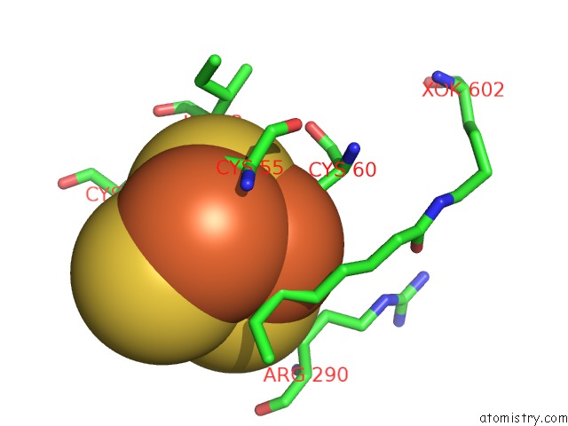
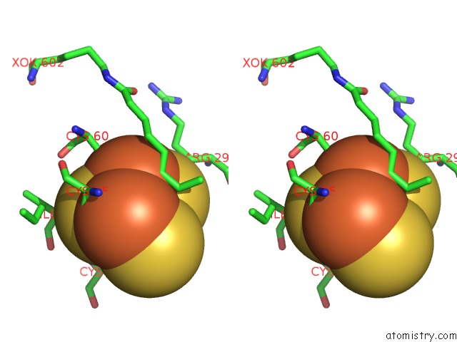
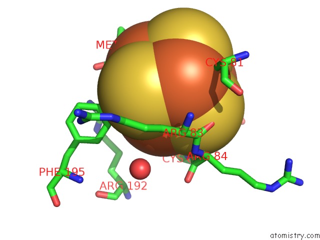
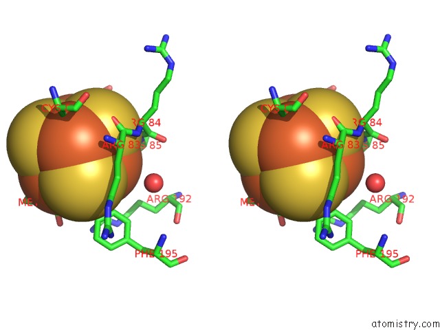
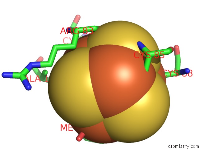
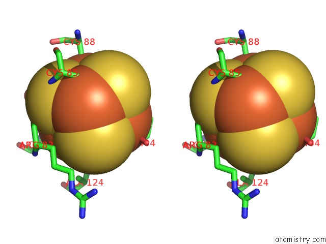
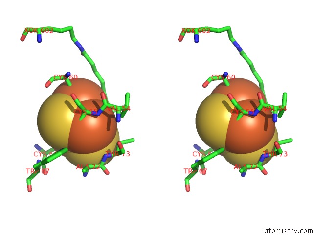
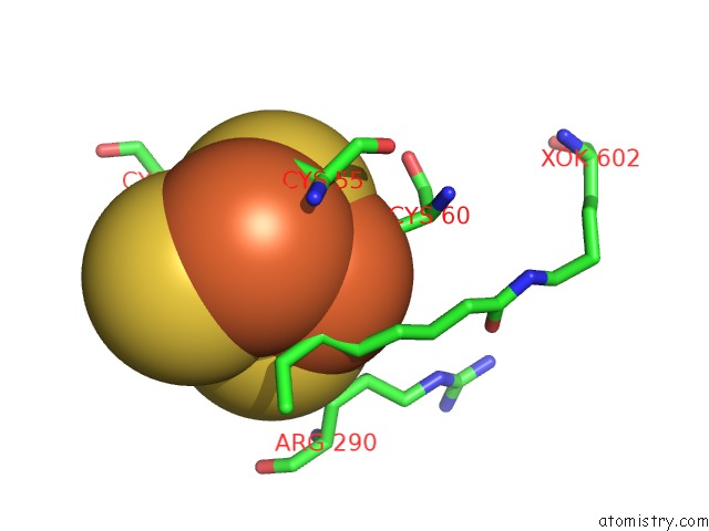
Iron binding site 1 out of 42 in 5exk
Go back to
Iron binding site 1 out
of 42 in the Crystal Structure of M. Tuberculosis Lipoyl Synthase with 6- Thiooctanoyl Peptide Intermediate
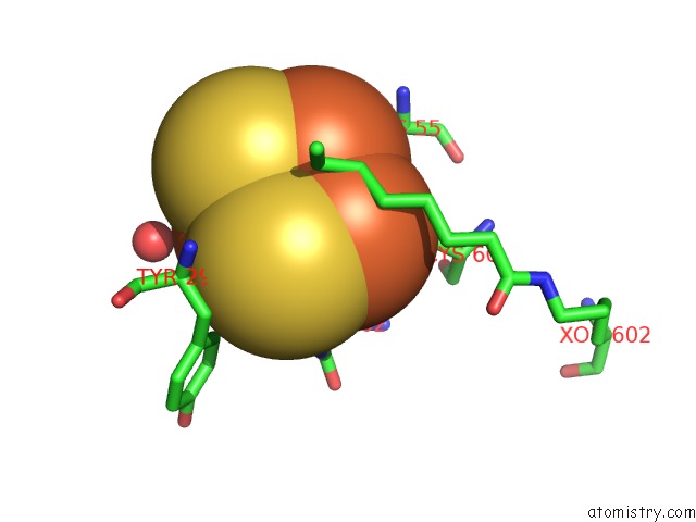
Mono view
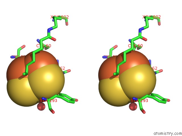
Stereo pair view
Mono view
Stereo pair view
A full contact list of Iron with other atoms in the Fe binding
site number 1 of Crystal Structure of M. Tuberculosis Lipoyl Synthase with 6- Thiooctanoyl Peptide Intermediate within 5.0Å range:
|
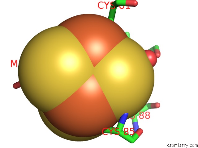
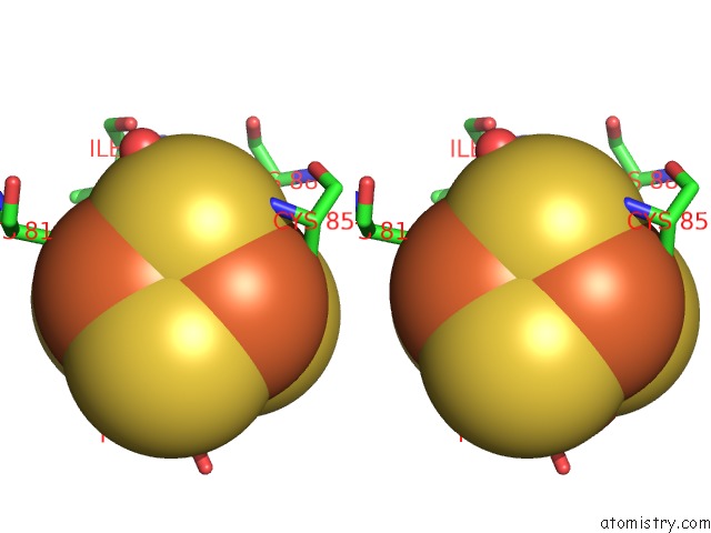
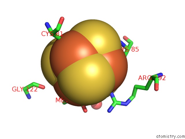
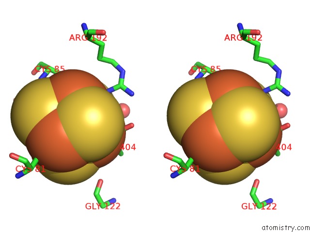
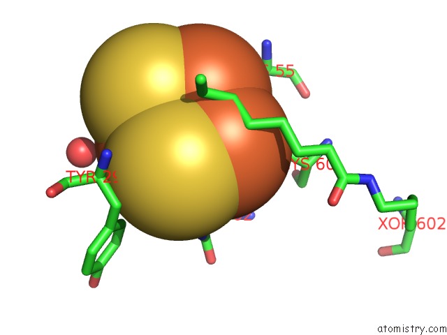
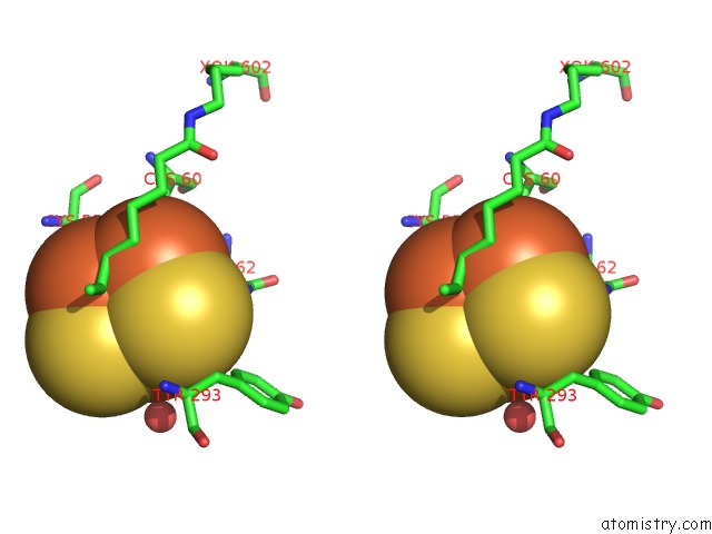
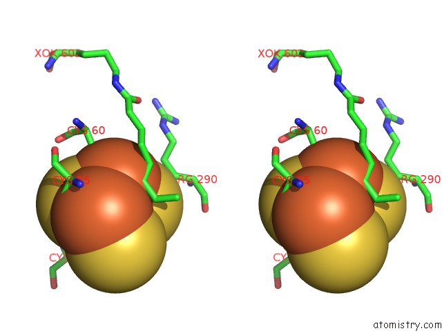
Iron binding site 2 out of 42 in 5exk
Go back to
Iron binding site 2 out
of 42 in the Crystal Structure of M. Tuberculosis Lipoyl Synthase with 6- Thiooctanoyl Peptide Intermediate
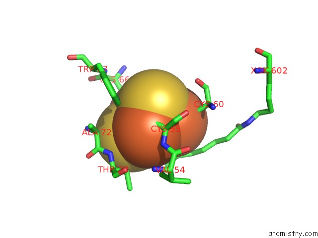
Mono view
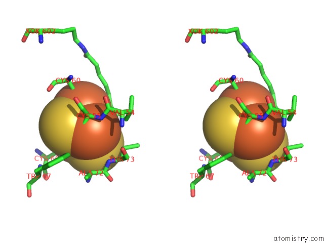
Stereo pair view
Mono view
Stereo pair view
A full contact list of Iron with other atoms in the Fe binding
site number 2 of Crystal Structure of M. Tuberculosis Lipoyl Synthase with 6- Thiooctanoyl Peptide Intermediate within 5.0Å range:
|
Iron binding site 3 out of 42 in 5exk
Go back to
Iron binding site 3 out
of 42 in the Crystal Structure of M. Tuberculosis Lipoyl Synthase with 6- Thiooctanoyl Peptide Intermediate
Mono view
Stereo pair view
Mono view
Stereo pair view
A full contact list of Iron with other atoms in the Fe binding
site number 3 of Crystal Structure of M. Tuberculosis Lipoyl Synthase with 6- Thiooctanoyl Peptide Intermediate within 5.0Å range:
|
Iron binding site 4 out of 42 in 5exk
Go back to
Iron binding site 4 out
of 42 in the Crystal Structure of M. Tuberculosis Lipoyl Synthase with 6- Thiooctanoyl Peptide Intermediate
Mono view
Stereo pair view
Mono view
Stereo pair view
A full contact list of Iron with other atoms in the Fe binding
site number 4 of Crystal Structure of M. Tuberculosis Lipoyl Synthase with 6- Thiooctanoyl Peptide Intermediate within 5.0Å range:
|
Iron binding site 5 out of 42 in 5exk
Go back to
Iron binding site 5 out
of 42 in the Crystal Structure of M. Tuberculosis Lipoyl Synthase with 6- Thiooctanoyl Peptide Intermediate
Mono view
Stereo pair view
Mono view
Stereo pair view
A full contact list of Iron with other atoms in the Fe binding
site number 5 of Crystal Structure of M. Tuberculosis Lipoyl Synthase with 6- Thiooctanoyl Peptide Intermediate within 5.0Å range:
|
Iron binding site 6 out of 42 in 5exk
Go back to
Iron binding site 6 out
of 42 in the Crystal Structure of M. Tuberculosis Lipoyl Synthase with 6- Thiooctanoyl Peptide Intermediate
Mono view
Stereo pair view
Mono view
Stereo pair view
A full contact list of Iron with other atoms in the Fe binding
site number 6 of Crystal Structure of M. Tuberculosis Lipoyl Synthase with 6- Thiooctanoyl Peptide Intermediate within 5.0Å range:
|
Iron binding site 7 out of 42 in 5exk
Go back to
Iron binding site 7 out
of 42 in the Crystal Structure of M. Tuberculosis Lipoyl Synthase with 6- Thiooctanoyl Peptide Intermediate
Mono view
Stereo pair view
Mono view
Stereo pair view
A full contact list of Iron with other atoms in the Fe binding
site number 7 of Crystal Structure of M. Tuberculosis Lipoyl Synthase with 6- Thiooctanoyl Peptide Intermediate within 5.0Å range:
|
Iron binding site 8 out of 42 in 5exk
Go back to
Iron binding site 8 out
of 42 in the Crystal Structure of M. Tuberculosis Lipoyl Synthase with 6- Thiooctanoyl Peptide Intermediate
Mono view
Stereo pair view
Mono view
Stereo pair view
A full contact list of Iron with other atoms in the Fe binding
site number 8 of Crystal Structure of M. Tuberculosis Lipoyl Synthase with 6- Thiooctanoyl Peptide Intermediate within 5.0Å range:
|
Iron binding site 9 out of 42 in 5exk
Go back to
Iron binding site 9 out
of 42 in the Crystal Structure of M. Tuberculosis Lipoyl Synthase with 6- Thiooctanoyl Peptide Intermediate

Mono view
Stereo pair view
Mono view
Stereo pair view
A full contact list of Iron with other atoms in the Fe binding
site number 9 of Crystal Structure of M. Tuberculosis Lipoyl Synthase with 6- Thiooctanoyl Peptide Intermediate within 5.0Å range:
|
Iron binding site 10 out of 42 in 5exk
Go back to
Iron binding site 10 out
of 42 in the Crystal Structure of M. Tuberculosis Lipoyl Synthase with 6- Thiooctanoyl Peptide Intermediate
Mono view
Stereo pair view
Mono view
Stereo pair view
A full contact list of Iron with other atoms in the Fe binding
site number 10 of Crystal Structure of M. Tuberculosis Lipoyl Synthase with 6- Thiooctanoyl Peptide Intermediate within 5.0Å range:
|
Reference:
M.I.Mclaughlin,
N.D.Lanz,
P.J.Goldman,
K.H.Lee,
S.J.Booker,
C.L.Drennan.
Crystallographic Snapshots of Sulfur Insertion By Lipoyl Synthase. Proc.Natl.Acad.Sci.Usa V. 113 9446 2016.
ISSN: ESSN 1091-6490
PubMed: 27506792
DOI: 10.1073/PNAS.1602486113
Page generated: Tue Aug 6 00:37:02 2024
ISSN: ESSN 1091-6490
PubMed: 27506792
DOI: 10.1073/PNAS.1602486113
Last articles
F in 7MLDF in 7MKX
F in 7MGK
F in 7MGJ
F in 7MHD
F in 7MFH
F in 7MHC
F in 7MGE
F in 7MFD
F in 7MEW