Iron »
PDB 5exd-5foi »
5eyj »
Iron in PDB 5eyj: Crystal Structure of Murine Neuroglobin Mutant V101F at 240 Mpa Pressure
Protein crystallography data
The structure of Crystal Structure of Murine Neuroglobin Mutant V101F at 240 Mpa Pressure, PDB code: 5eyj
was solved by
N.Colloc'h,
E.Girard,
B.Vallone,
T.Prange,
with X-Ray Crystallography technique. A brief refinement statistics is given in the table below:
| Resolution Low / High (Å) | 20.00 / 2.40 |
| Space group | H 3 2 |
| Cell size a, b, c (Å), α, β, γ (°) | 87.199, 87.199, 113.904, 90.00, 90.00, 120.00 |
| R / Rfree (%) | 14.4 / 26 |
Iron Binding Sites:
The binding sites of Iron atom in the Crystal Structure of Murine Neuroglobin Mutant V101F at 240 Mpa Pressure
(pdb code 5eyj). This binding sites where shown within
5.0 Angstroms radius around Iron atom.
In total only one binding site of Iron was determined in the Crystal Structure of Murine Neuroglobin Mutant V101F at 240 Mpa Pressure, PDB code: 5eyj:
In total only one binding site of Iron was determined in the Crystal Structure of Murine Neuroglobin Mutant V101F at 240 Mpa Pressure, PDB code: 5eyj:
Iron binding site 1 out of 1 in 5eyj
Go back to
Iron binding site 1 out
of 1 in the Crystal Structure of Murine Neuroglobin Mutant V101F at 240 Mpa Pressure
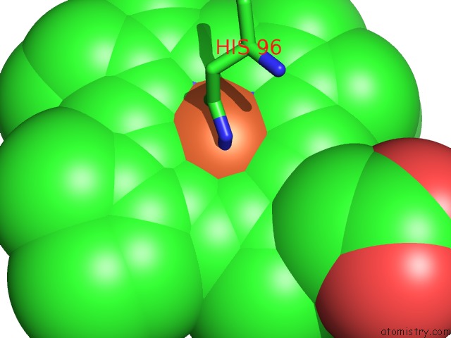
Mono view
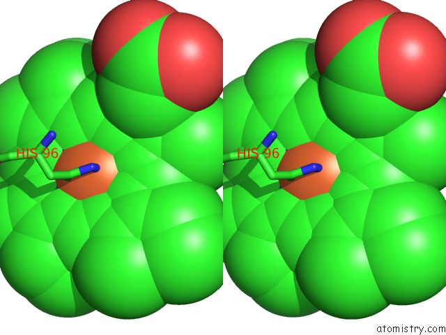
Stereo pair view
Mono view
Stereo pair view
A full contact list of Iron with other atoms in the Fe binding
site number 1 of Crystal Structure of Murine Neuroglobin Mutant V101F at 240 Mpa Pressure within 5.0Å range:
|
Reference:
N.Colloc'h,
S.Sacquin-Mora,
G.Avella,
A.C.Dhaussy,
T.Prange,
B.Vallone,
E.Girard.
Determinants of Neuroglobin Plasticity Highlighted By Joint Coarse-Grained Simulations and High Pressure Crystallography. Sci Rep V. 7 1858 2017.
ISSN: ESSN 2045-2322
PubMed: 28500341
DOI: 10.1038/S41598-017-02097-1
Page generated: Tue Aug 6 00:37:01 2024
ISSN: ESSN 2045-2322
PubMed: 28500341
DOI: 10.1038/S41598-017-02097-1
Last articles
Fe in 2YXOFe in 2YRS
Fe in 2YXC
Fe in 2YNM
Fe in 2YVJ
Fe in 2YP1
Fe in 2YU2
Fe in 2YU1
Fe in 2YQB
Fe in 2YOO