Iron »
PDB 5exd-5foi »
5fne »
Iron in PDB 5fne: Crystal Structure of Fungal Versatile Peroxidase From Pleurotus Eryngii Triple Mutant E37K, H39R & G330R
Enzymatic activity of Crystal Structure of Fungal Versatile Peroxidase From Pleurotus Eryngii Triple Mutant E37K, H39R & G330R
All present enzymatic activity of Crystal Structure of Fungal Versatile Peroxidase From Pleurotus Eryngii Triple Mutant E37K, H39R & G330R:
1.11.1.16;
1.11.1.16;
Protein crystallography data
The structure of Crystal Structure of Fungal Versatile Peroxidase From Pleurotus Eryngii Triple Mutant E37K, H39R & G330R, PDB code: 5fne
was solved by
F.J.Medrano,
A.Romero,
with X-Ray Crystallography technique. A brief refinement statistics is given in the table below:
| Resolution Low / High (Å) | 44.778 / 1.50 |
| Space group | P 21 21 2 |
| Cell size a, b, c (Å), α, β, γ (°) | 55.190, 104.200, 76.600, 90.00, 90.00, 90.00 |
| R / Rfree (%) | 19.24 / 22.92 |
Other elements in 5fne:
The structure of Crystal Structure of Fungal Versatile Peroxidase From Pleurotus Eryngii Triple Mutant E37K, H39R & G330R also contains other interesting chemical elements:
| Calcium | (Ca) | 2 atoms |
Iron Binding Sites:
The binding sites of Iron atom in the Crystal Structure of Fungal Versatile Peroxidase From Pleurotus Eryngii Triple Mutant E37K, H39R & G330R
(pdb code 5fne). This binding sites where shown within
5.0 Angstroms radius around Iron atom.
In total only one binding site of Iron was determined in the Crystal Structure of Fungal Versatile Peroxidase From Pleurotus Eryngii Triple Mutant E37K, H39R & G330R, PDB code: 5fne:
In total only one binding site of Iron was determined in the Crystal Structure of Fungal Versatile Peroxidase From Pleurotus Eryngii Triple Mutant E37K, H39R & G330R, PDB code: 5fne:
Iron binding site 1 out of 1 in 5fne
Go back to
Iron binding site 1 out
of 1 in the Crystal Structure of Fungal Versatile Peroxidase From Pleurotus Eryngii Triple Mutant E37K, H39R & G330R
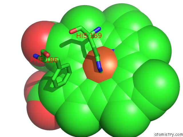
Mono view
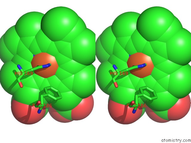
Stereo pair view
Mono view
Stereo pair view
A full contact list of Iron with other atoms in the Fe binding
site number 1 of Crystal Structure of Fungal Versatile Peroxidase From Pleurotus Eryngii Triple Mutant E37K, H39R & G330R within 5.0Å range:
|
Reference:
V.Saez-Jimenez,
S.Acebes,
E.Garcia-Ruiz,
A.Romero,
V.Guallar,
M.Alcalde,
F.J.Medrano,
A.T.Martinez,
F.J.Ruiz-Duenas.
Unveiling the Basis of Alkaline Stability of An Evolved Versatile Peroxidase. Biochem.J. V. 473 1917 2016.
ISSN: ISSN 0264-6021
PubMed: 27118867
DOI: 10.1042/BCJ20160248
Page generated: Tue Aug 6 00:59:55 2024
ISSN: ISSN 0264-6021
PubMed: 27118867
DOI: 10.1042/BCJ20160248
Last articles
Zn in 9J0NZn in 9J0O
Zn in 9J0P
Zn in 9FJX
Zn in 9EKB
Zn in 9C0F
Zn in 9CAH
Zn in 9CH0
Zn in 9CH3
Zn in 9CH1