Iron »
PDB 5h92-5ibe »
5hav »
Iron in PDB 5hav: Sperm Whale Myoglobin Mutant L29H F33Y F43H (F33Y Cubmb) with Oxygen Bound
Protein crystallography data
The structure of Sperm Whale Myoglobin Mutant L29H F33Y F43H (F33Y Cubmb) with Oxygen Bound, PDB code: 5hav
was solved by
I.D.Petrik,
Y.Lu,
with X-Ray Crystallography technique. A brief refinement statistics is given in the table below:
| Resolution Low / High (Å) | 23.83 / 1.27 |
| Space group | P 21 21 21 |
| Cell size a, b, c (Å), α, β, γ (°) | 39.632, 47.609, 76.534, 90.00, 90.00, 90.00 |
| R / Rfree (%) | 14.6 / 17.8 |
Iron Binding Sites:
The binding sites of Iron atom in the Sperm Whale Myoglobin Mutant L29H F33Y F43H (F33Y Cubmb) with Oxygen Bound
(pdb code 5hav). This binding sites where shown within
5.0 Angstroms radius around Iron atom.
In total only one binding site of Iron was determined in the Sperm Whale Myoglobin Mutant L29H F33Y F43H (F33Y Cubmb) with Oxygen Bound, PDB code: 5hav:
In total only one binding site of Iron was determined in the Sperm Whale Myoglobin Mutant L29H F33Y F43H (F33Y Cubmb) with Oxygen Bound, PDB code: 5hav:
Iron binding site 1 out of 1 in 5hav
Go back to
Iron binding site 1 out
of 1 in the Sperm Whale Myoglobin Mutant L29H F33Y F43H (F33Y Cubmb) with Oxygen Bound
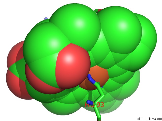
Mono view
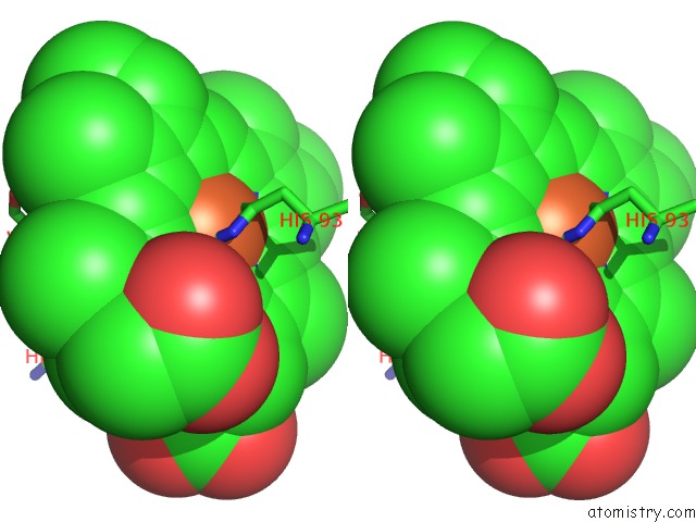
Stereo pair view
Mono view
Stereo pair view
A full contact list of Iron with other atoms in the Fe binding
site number 1 of Sperm Whale Myoglobin Mutant L29H F33Y F43H (F33Y Cubmb) with Oxygen Bound within 5.0Å range:
|
Reference:
I.D.Petrik,
R.Davydov,
M.Ross,
X.Zhao,
B.Hoffman,
Y.Lu.
Spectroscopic and Crystallographic Evidence For the Role of A Water-Containing H-Bond Network in Oxidase Activity of An Engineered Myoglobin. J.Am.Chem.Soc. V. 138 1134 2016.
ISSN: ESSN 1520-5126
PubMed: 26716352
DOI: 10.1021/JACS.5B12004
Page generated: Tue Aug 6 01:52:32 2024
ISSN: ESSN 1520-5126
PubMed: 26716352
DOI: 10.1021/JACS.5B12004
Last articles
Fe in 2YXOFe in 2YRS
Fe in 2YXC
Fe in 2YNM
Fe in 2YVJ
Fe in 2YP1
Fe in 2YU2
Fe in 2YU1
Fe in 2YQB
Fe in 2YOO