Iron »
PDB 5h92-5ibe »
5hio »
Iron in PDB 5hio: Crystal Structure of Pqs Response Protein Pqse in Complex with 2- Aminobenzoylacetate
Protein crystallography data
The structure of Crystal Structure of Pqs Response Protein Pqse in Complex with 2- Aminobenzoylacetate, PDB code: 5hio
was solved by
F.Witzgall,
W.Blankenfeldt,
with X-Ray Crystallography technique. A brief refinement statistics is given in the table below:
| Resolution Low / High (Å) | 48.66 / 1.90 |
| Space group | P 32 2 1 |
| Cell size a, b, c (Å), α, β, γ (°) | 61.124, 61.124, 145.979, 90.00, 90.00, 120.00 |
| R / Rfree (%) | 16.4 / 19.8 |
Iron Binding Sites:
The binding sites of Iron atom in the Crystal Structure of Pqs Response Protein Pqse in Complex with 2- Aminobenzoylacetate
(pdb code 5hio). This binding sites where shown within
5.0 Angstroms radius around Iron atom.
In total 2 binding sites of Iron where determined in the Crystal Structure of Pqs Response Protein Pqse in Complex with 2- Aminobenzoylacetate, PDB code: 5hio:
Jump to Iron binding site number: 1; 2;
In total 2 binding sites of Iron where determined in the Crystal Structure of Pqs Response Protein Pqse in Complex with 2- Aminobenzoylacetate, PDB code: 5hio:
Jump to Iron binding site number: 1; 2;
Iron binding site 1 out of 2 in 5hio
Go back to
Iron binding site 1 out
of 2 in the Crystal Structure of Pqs Response Protein Pqse in Complex with 2- Aminobenzoylacetate
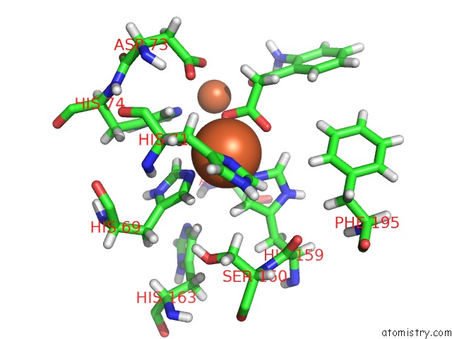
Mono view
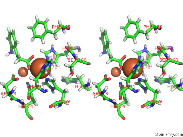
Stereo pair view
Mono view
Stereo pair view
A full contact list of Iron with other atoms in the Fe binding
site number 1 of Crystal Structure of Pqs Response Protein Pqse in Complex with 2- Aminobenzoylacetate within 5.0Å range:
|
Iron binding site 2 out of 2 in 5hio
Go back to
Iron binding site 2 out
of 2 in the Crystal Structure of Pqs Response Protein Pqse in Complex with 2- Aminobenzoylacetate
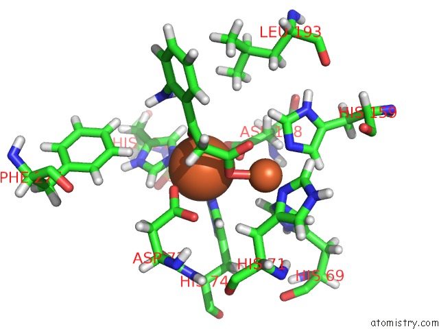
Mono view
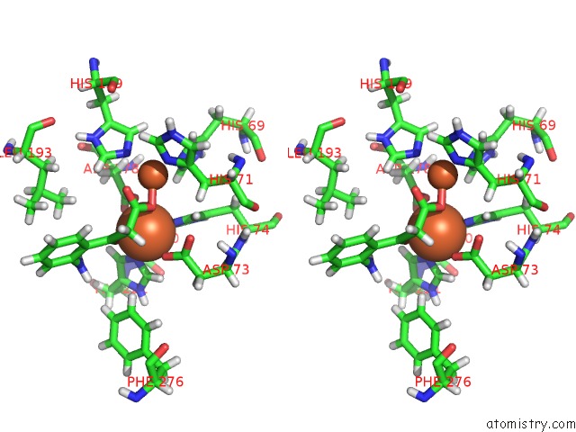
Stereo pair view
Mono view
Stereo pair view
A full contact list of Iron with other atoms in the Fe binding
site number 2 of Crystal Structure of Pqs Response Protein Pqse in Complex with 2- Aminobenzoylacetate within 5.0Å range:
|
Reference:
M.Zender,
F.Witzgall,
S.L.Drees,
E.Weidel,
C.K.Maurer,
S.Fetzner,
W.Blankenfeldt,
M.Empting,
R.W.Hartmann.
Dissecting the Multiple Roles of Pqse in Pseudomonas Aeruginosa Virulence By Discovery of Small Tool Compounds. Acs Chem.Biol. V. 11 1755 2016.
ISSN: ESSN 1554-8937
PubMed: 27082157
DOI: 10.1021/ACSCHEMBIO.6B00156
Page generated: Tue Aug 6 01:54:03 2024
ISSN: ESSN 1554-8937
PubMed: 27082157
DOI: 10.1021/ACSCHEMBIO.6B00156
Last articles
Zn in 9J0NZn in 9J0O
Zn in 9J0P
Zn in 9FJX
Zn in 9EKB
Zn in 9C0F
Zn in 9CAH
Zn in 9CH0
Zn in 9CH3
Zn in 9CH1