Iron in PDB 5his: Crystal Structure of Pqs Response Protein Pqse in Complex with 3- Methylthiophene-2-Carboxylic Acid
Protein crystallography data
The structure of Crystal Structure of Pqs Response Protein Pqse in Complex with 3- Methylthiophene-2-Carboxylic Acid, PDB code: 5his
was solved by
F.Witzgall,
W.Blankenfeldt,
with X-Ray Crystallography technique. A brief refinement statistics is given in the table below:
Resolution Low / High (Å)
|
48.76 /
1.77
|
Space group
|
P 32 2 1
|
Cell size a, b, c (Å), α, β, γ (°)
|
61.144,
61.144,
146.277,
90.00,
90.00,
120.00
|
R / Rfree (%)
|
16.2 /
20
|
Iron Binding Sites:
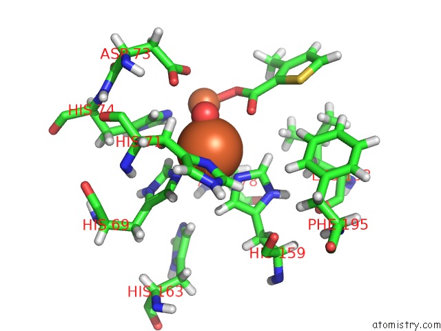
The binding sites of Iron atom in the Crystal Structure of Pqs Response Protein Pqse in Complex with 3- Methylthiophene-2-Carboxylic Acid
(pdb code 5his). This binding sites where shown within
5.0 Angstroms radius around Iron atom.
In total 2 binding sites of Iron where determined in the
Crystal Structure of Pqs Response Protein Pqse in Complex with 3- Methylthiophene-2-Carboxylic Acid, PDB code: 5his:
Jump to Iron binding site number:
1;
2;
Iron binding site 1 out
of 2 in 5his
Go back to
Iron Binding Sites List in 5his
Iron binding site 1 out
of 2 in the Crystal Structure of Pqs Response Protein Pqse in Complex with 3- Methylthiophene-2-Carboxylic Acid
Mono view
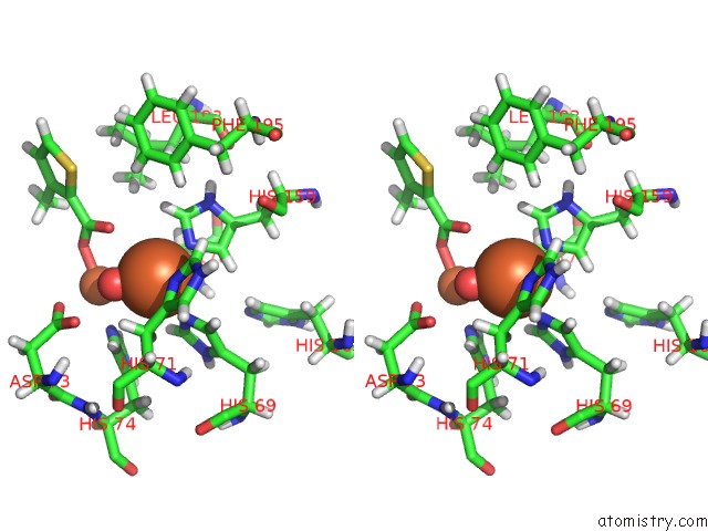
Stereo pair view
|
A full contact list of Iron with other atoms in the Fe binding
site number 1 of Crystal Structure of Pqs Response Protein Pqse in Complex with 3- Methylthiophene-2-Carboxylic Acid within 5.0Å range:
probe
|
atom
|
residue
|
distance (Å)
|
B
|
Occ
|
A:Fe401
b:27.8
occ:1.00
|
O
|
A:HOH520
|
1.9
|
22.8
|
1.0
|
NE2
|
A:HIS159
|
2.1
|
26.0
|
1.0
|
OD2
|
A:ASP178
|
2.2
|
21.8
|
1.0
|
NE2
|
A:HIS69
|
2.2
|
23.9
|
1.0
|
ND1
|
A:HIS71
|
2.6
|
26.3
|
1.0
|
O09
|
A:60P403
|
3.1
|
39.0
|
1.0
|
CE1
|
A:HIS159
|
3.1
|
25.1
|
1.0
|
CE1
|
A:HIS69
|
3.1
|
23.0
|
1.0
|
FE
|
A:FE402
|
3.1
|
25.9
|
1.0
|
HB2
|
A:HIS71
|
3.1
|
32.9
|
1.0
|
CD2
|
A:HIS159
|
3.2
|
25.4
|
1.0
|
HE1
|
A:HIS69
|
3.2
|
27.6
|
1.0
|
CD2
|
A:HIS69
|
3.2
|
23.3
|
1.0
|
HE1
|
A:HIS159
|
3.2
|
30.2
|
1.0
|
CG
|
A:ASP178
|
3.3
|
26.0
|
1.0
|
HB2
|
A:ASP178
|
3.3
|
27.4
|
1.0
|
HD2
|
A:HIS159
|
3.3
|
30.5
|
1.0
|
HD2
|
A:HIS69
|
3.4
|
27.9
|
1.0
|
CG
|
A:HIS71
|
3.5
|
29.8
|
1.0
|
CE1
|
A:HIS71
|
3.5
|
33.7
|
1.0
|
HD2
|
A:HIS74
|
3.5
|
27.8
|
1.0
|
CB
|
A:HIS71
|
3.6
|
27.4
|
1.0
|
HE1
|
A:HIS71
|
3.7
|
40.4
|
1.0
|
HB3
|
A:HIS71
|
3.7
|
32.9
|
1.0
|
CB
|
A:ASP178
|
3.8
|
22.9
|
1.0
|
NE2
|
A:HIS74
|
3.8
|
23.5
|
1.0
|
CD2
|
A:HIS74
|
3.8
|
23.1
|
1.0
|
O08
|
A:60P403
|
3.9
|
26.3
|
1.0
|
C07
|
A:60P403
|
3.9
|
33.9
|
1.0
|
HB3
|
A:ASP178
|
4.0
|
27.4
|
1.0
|
ND1
|
A:HIS159
|
4.2
|
26.1
|
1.0
|
OD1
|
A:ASP73
|
4.2
|
27.1
|
1.0
|
ND1
|
A:HIS69
|
4.2
|
21.3
|
1.0
|
CG
|
A:HIS159
|
4.3
|
24.9
|
1.0
|
CG
|
A:HIS69
|
4.3
|
22.7
|
1.0
|
OD1
|
A:ASP178
|
4.4
|
26.0
|
1.0
|
OD2
|
A:ASP73
|
4.6
|
27.4
|
1.0
|
CD2
|
A:HIS71
|
4.6
|
31.9
|
1.0
|
NE2
|
A:HIS71
|
4.6
|
34.4
|
1.0
|
HD2
|
A:HIS163
|
4.7
|
30.1
|
1.0
|
HD2
|
A:PHE195
|
4.8
|
37.2
|
1.0
|
CE1
|
A:HIS74
|
4.8
|
24.7
|
1.0
|
CG
|
A:HIS74
|
4.8
|
25.0
|
1.0
|
CG
|
A:ASP73
|
4.8
|
28.9
|
1.0
|
H
|
A:HIS71
|
4.9
|
32.6
|
1.0
|
HD1
|
A:HIS159
|
5.0
|
31.3
|
1.0
|
HD13
|
A:LEU193
|
5.0
|
30.3
|
1.0
|
HE2
|
A:PHE195
|
5.0
|
36.8
|
1.0
|
|
Iron binding site 2 out
of 2 in 5his
Go back to
Iron Binding Sites List in 5his
Iron binding site 2 out
of 2 in the Crystal Structure of Pqs Response Protein Pqse in Complex with 3- Methylthiophene-2-Carboxylic Acid
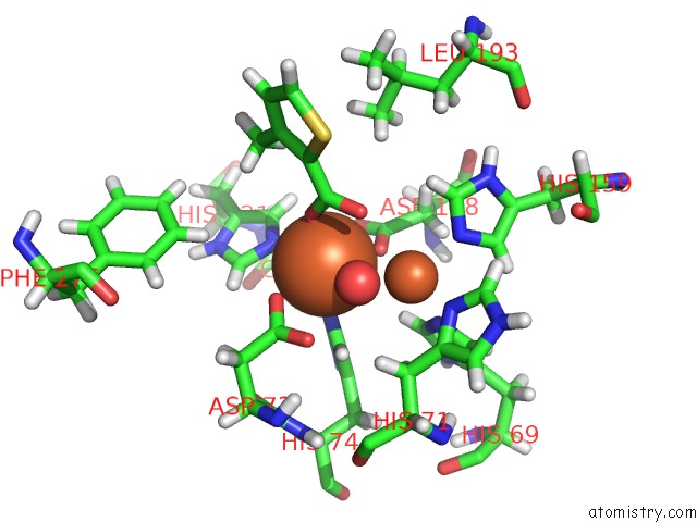
Mono view
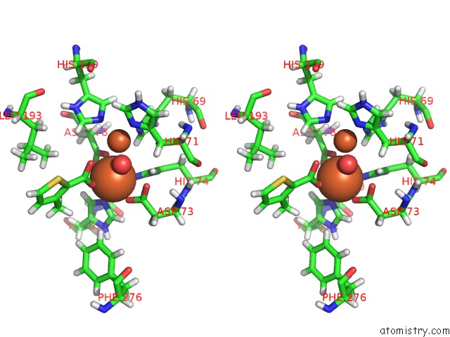
Stereo pair view
|
A full contact list of Iron with other atoms in the Fe binding
site number 2 of Crystal Structure of Pqs Response Protein Pqse in Complex with 3- Methylthiophene-2-Carboxylic Acid within 5.0Å range:
probe
|
atom
|
residue
|
distance (Å)
|
B
|
Occ
|
A:Fe402
b:25.9
occ:1.00
|
O08
|
A:60P403
|
2.0
|
26.3
|
1.0
|
O
|
A:HOH520
|
2.0
|
22.8
|
1.0
|
OD2
|
A:ASP178
|
2.1
|
21.8
|
1.0
|
NE2
|
A:HIS74
|
2.1
|
23.5
|
1.0
|
OD2
|
A:ASP73
|
2.3
|
27.4
|
1.0
|
NE2
|
A:HIS221
|
2.4
|
23.8
|
1.0
|
CG
|
A:ASP178
|
2.9
|
26.0
|
1.0
|
C07
|
A:60P403
|
3.0
|
33.9
|
1.0
|
CD2
|
A:HIS74
|
3.0
|
23.1
|
1.0
|
FE
|
A:FE401
|
3.1
|
27.8
|
1.0
|
OD1
|
A:ASP178
|
3.1
|
26.0
|
1.0
|
HD2
|
A:HIS74
|
3.1
|
27.8
|
1.0
|
CE1
|
A:HIS74
|
3.2
|
24.7
|
1.0
|
O09
|
A:60P403
|
3.2
|
39.0
|
1.0
|
CG
|
A:ASP73
|
3.2
|
28.9
|
1.0
|
CE1
|
A:HIS221
|
3.3
|
24.7
|
1.0
|
HE1
|
A:HIS221
|
3.3
|
29.7
|
1.0
|
CD2
|
A:HIS221
|
3.4
|
23.0
|
1.0
|
HE1
|
A:HIS74
|
3.5
|
29.6
|
1.0
|
OD1
|
A:ASP73
|
3.6
|
27.1
|
1.0
|
HD2
|
A:HIS221
|
3.6
|
27.6
|
1.0
|
HE1
|
A:HIS69
|
3.8
|
27.6
|
1.0
|
CG
|
A:HIS74
|
4.2
|
25.0
|
1.0
|
H063
|
A:60P403
|
4.3
|
33.5
|
1.0
|
ND1
|
A:HIS74
|
4.3
|
23.7
|
1.0
|
C02
|
A:60P403
|
4.3
|
32.9
|
1.0
|
CB
|
A:ASP178
|
4.3
|
22.9
|
1.0
|
HE1
|
A:PHE276
|
4.4
|
35.4
|
1.0
|
NE2
|
A:HIS69
|
4.4
|
23.9
|
1.0
|
CE1
|
A:HIS69
|
4.4
|
23.0
|
1.0
|
H062
|
A:60P403
|
4.4
|
33.5
|
1.0
|
ND1
|
A:HIS221
|
4.4
|
24.6
|
1.0
|
CG
|
A:HIS221
|
4.5
|
23.3
|
1.0
|
HB2
|
A:ASP73
|
4.5
|
32.0
|
1.0
|
HB2
|
A:ASP178
|
4.6
|
27.4
|
1.0
|
CB
|
A:ASP73
|
4.6
|
26.6
|
1.0
|
HB3
|
A:ASP178
|
4.6
|
27.4
|
1.0
|
C06
|
A:60P403
|
4.7
|
28.0
|
1.0
|
NE2
|
A:HIS159
|
4.8
|
26.0
|
1.0
|
HE1
|
A:HIS159
|
4.8
|
30.2
|
1.0
|
HD13
|
A:LEU193
|
4.8
|
30.3
|
1.0
|
HB2
|
A:HIS71
|
4.8
|
32.9
|
1.0
|
HA3
|
A:GLY220
|
4.8
|
29.1
|
1.0
|
HB3
|
A:HIS71
|
5.0
|
32.9
|
1.0
|
|
Reference:
M.Zender,
F.Witzgall,
S.L.Drees,
E.Weidel,
C.K.Maurer,
S.Fetzner,
W.Blankenfeldt,
M.Empting,
R.W.Hartmann.
Dissecting the Multiple Roles of Pqse in Pseudomonas Aeruginosa Virulence By Discovery of Small Tool Compounds. Acs Chem.Biol. V. 11 1755 2016.
ISSN: ESSN 1554-8937
PubMed: 27082157
DOI: 10.1021/ACSCHEMBIO.6B00156
Page generated: Tue Aug 6 01:55:39 2024
|