Iron »
PDB 5h92-5ibe »
5hlu »
Iron in PDB 5hlu: X-Ray Crystal Structure of Met F43H/H64A Sperm Whale Myoglobin in Complex with Nitric Oxide
Protein crystallography data
The structure of X-Ray Crystal Structure of Met F43H/H64A Sperm Whale Myoglobin in Complex with Nitric Oxide, PDB code: 5hlu
was solved by
H.Yuan,
with X-Ray Crystallography technique. A brief refinement statistics is given in the table below:
| Resolution Low / High (Å) | 41.75 / 1.50 |
| Space group | P 21 21 21 |
| Cell size a, b, c (Å), α, β, γ (°) | 39.690, 49.528, 77.598, 90.00, 90.00, 90.00 |
| R / Rfree (%) | 14.1 / 15.9 |
Iron Binding Sites:
The binding sites of Iron atom in the X-Ray Crystal Structure of Met F43H/H64A Sperm Whale Myoglobin in Complex with Nitric Oxide
(pdb code 5hlu). This binding sites where shown within
5.0 Angstroms radius around Iron atom.
In total only one binding site of Iron was determined in the X-Ray Crystal Structure of Met F43H/H64A Sperm Whale Myoglobin in Complex with Nitric Oxide, PDB code: 5hlu:
In total only one binding site of Iron was determined in the X-Ray Crystal Structure of Met F43H/H64A Sperm Whale Myoglobin in Complex with Nitric Oxide, PDB code: 5hlu:
Iron binding site 1 out of 1 in 5hlu
Go back to
Iron binding site 1 out
of 1 in the X-Ray Crystal Structure of Met F43H/H64A Sperm Whale Myoglobin in Complex with Nitric Oxide
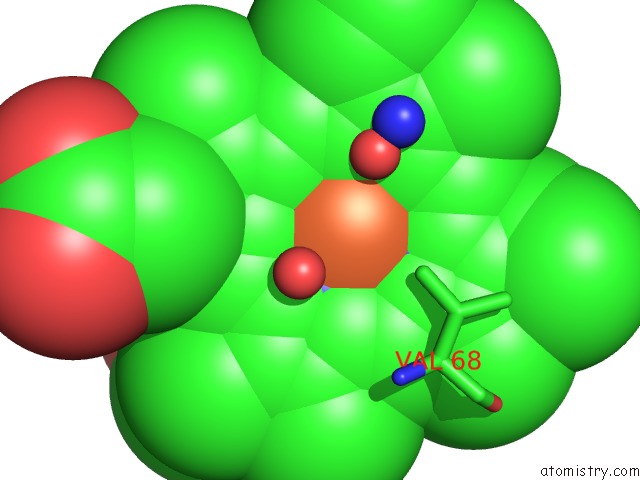
Mono view
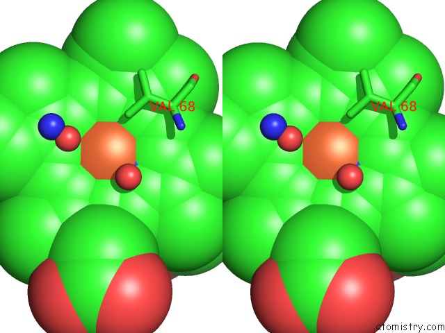
Stereo pair view
Mono view
Stereo pair view
A full contact list of Iron with other atoms in the Fe binding
site number 1 of X-Ray Crystal Structure of Met F43H/H64A Sperm Whale Myoglobin in Complex with Nitric Oxide within 5.0Å range:
|
Reference:
L.B.Wu,
H.Yuan,
S.Q.Gao,
Y.You,
C.M.Nie,
G.B.Wen,
Y.W.Lin,
X.Tan.
Regulating the Nitrite Reductase Activity of Myoglobin By Redesigning the Heme Active Center Nitric Oxide V. 57 21 2016.
ISSN: ESSN 1089-8611
PubMed: 27108710
DOI: 10.1016/J.NIOX.2016.04.007
Page generated: Tue Aug 6 01:57:21 2024
ISSN: ESSN 1089-8611
PubMed: 27108710
DOI: 10.1016/J.NIOX.2016.04.007
Last articles
Zn in 9J0NZn in 9J0O
Zn in 9J0P
Zn in 9FJX
Zn in 9EKB
Zn in 9C0F
Zn in 9CAH
Zn in 9CH0
Zn in 9CH3
Zn in 9CH1