Iron »
PDB 5h92-5ibe »
5hr6 »
Iron in PDB 5hr6: X-Ray Crystal Structure of C118A Rlmn with Cross-Linked Trna Purified From Escherichia Coli
Protein crystallography data
The structure of X-Ray Crystal Structure of C118A Rlmn with Cross-Linked Trna Purified From Escherichia Coli, PDB code: 5hr6
was solved by
E.L.Schwalm,
T.L.Grove,
S.J.Booker,
A.K.Boal,
with X-Ray Crystallography technique. A brief refinement statistics is given in the table below:
| Resolution Low / High (Å) | 88.66 / 2.88 |
| Space group | P 1 21 1 |
| Cell size a, b, c (Å), α, β, γ (°) | 88.664, 69.821, 149.298, 90.00, 90.30, 90.00 |
| R / Rfree (%) | 18.2 / 22.8 |
Other elements in 5hr6:
The structure of X-Ray Crystal Structure of C118A Rlmn with Cross-Linked Trna Purified From Escherichia Coli also contains other interesting chemical elements:
| Magnesium | (Mg) | 6 atoms |
Iron Binding Sites:
The binding sites of Iron atom in the X-Ray Crystal Structure of C118A Rlmn with Cross-Linked Trna Purified From Escherichia Coli
(pdb code 5hr6). This binding sites where shown within
5.0 Angstroms radius around Iron atom.
In total 8 binding sites of Iron where determined in the X-Ray Crystal Structure of C118A Rlmn with Cross-Linked Trna Purified From Escherichia Coli, PDB code: 5hr6:
Jump to Iron binding site number: 1; 2; 3; 4; 5; 6; 7; 8;
In total 8 binding sites of Iron where determined in the X-Ray Crystal Structure of C118A Rlmn with Cross-Linked Trna Purified From Escherichia Coli, PDB code: 5hr6:
Jump to Iron binding site number: 1; 2; 3; 4; 5; 6; 7; 8;
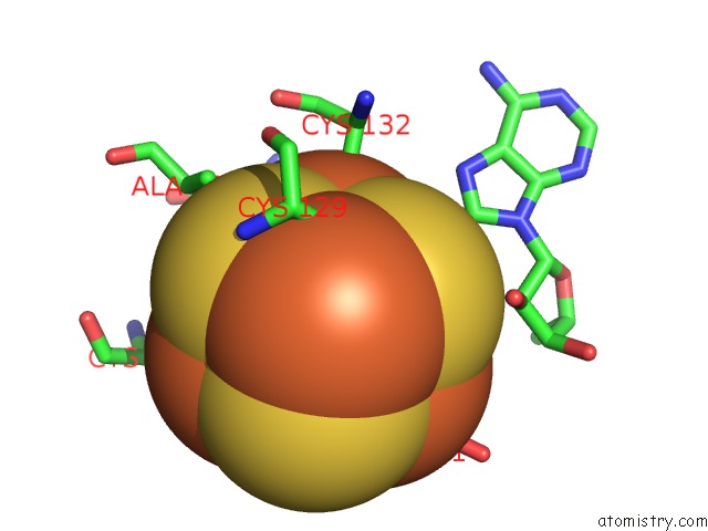
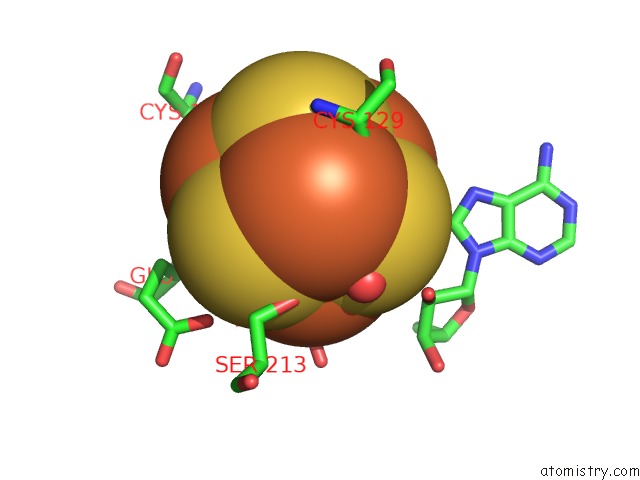
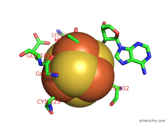
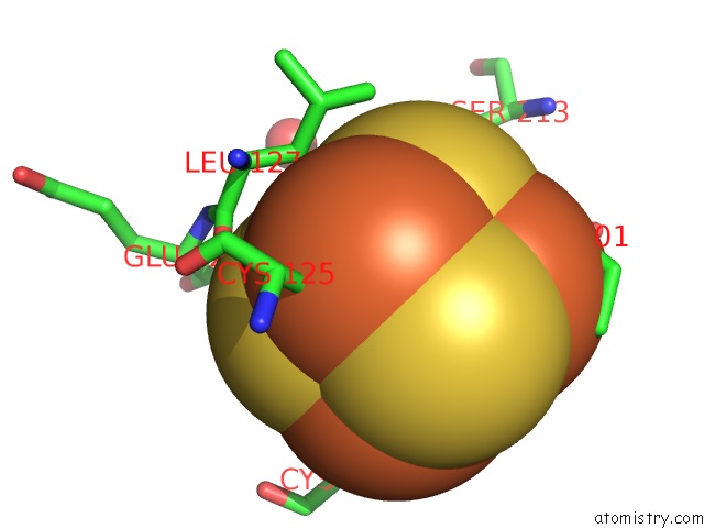
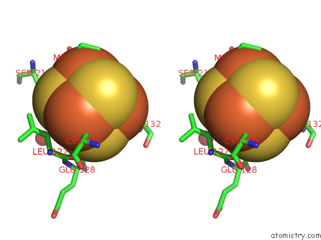
Iron binding site 1 out of 8 in 5hr6
Go back to
Iron binding site 1 out
of 8 in the X-Ray Crystal Structure of C118A Rlmn with Cross-Linked Trna Purified From Escherichia Coli
Mono view
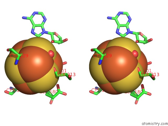
Stereo pair view
Mono view
Stereo pair view
A full contact list of Iron with other atoms in the Fe binding
site number 1 of X-Ray Crystal Structure of C118A Rlmn with Cross-Linked Trna Purified From Escherichia Coli within 5.0Å range:
|
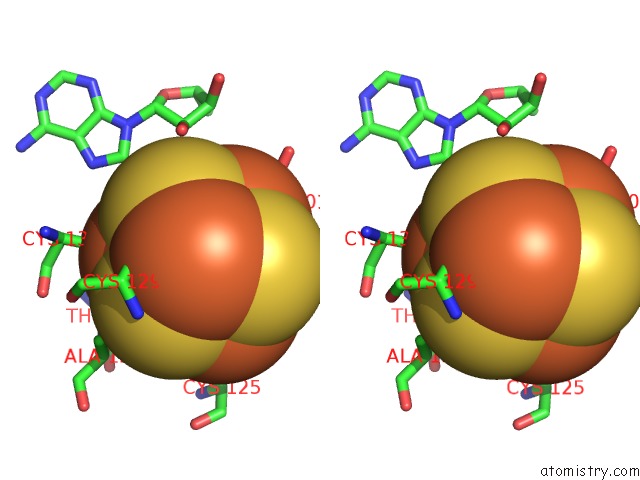
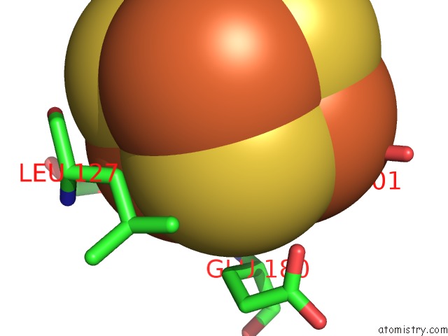
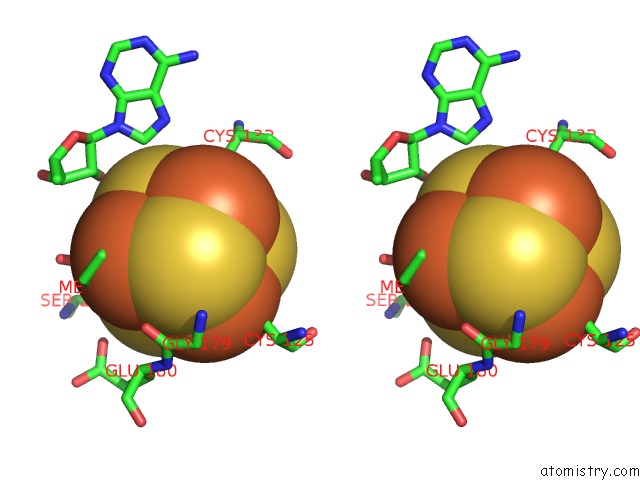
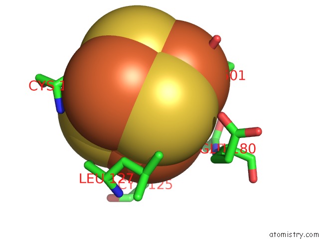
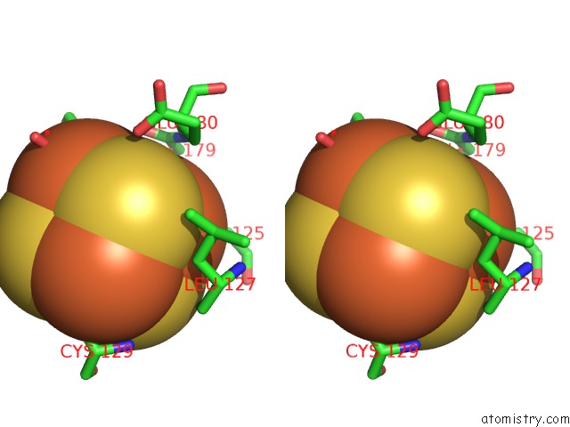
Iron binding site 2 out of 8 in 5hr6
Go back to
Iron binding site 2 out
of 8 in the X-Ray Crystal Structure of C118A Rlmn with Cross-Linked Trna Purified From Escherichia Coli
Mono view
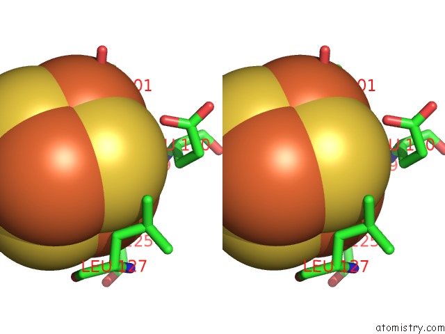
Stereo pair view
Mono view
Stereo pair view
A full contact list of Iron with other atoms in the Fe binding
site number 2 of X-Ray Crystal Structure of C118A Rlmn with Cross-Linked Trna Purified From Escherichia Coli within 5.0Å range:
|
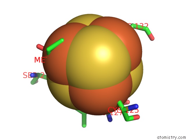
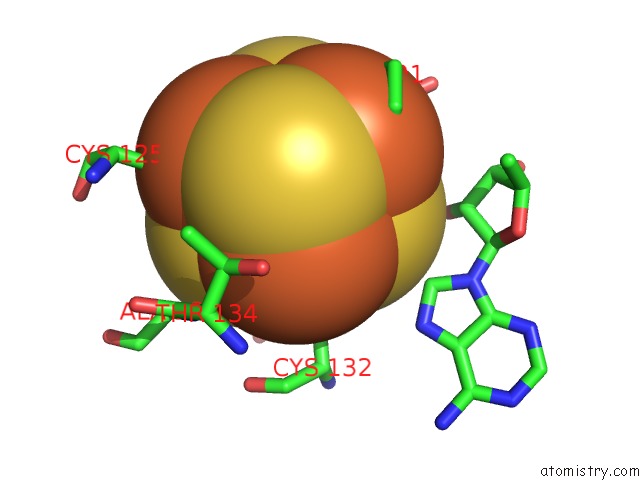
Iron binding site 3 out of 8 in 5hr6
Go back to
Iron binding site 3 out
of 8 in the X-Ray Crystal Structure of C118A Rlmn with Cross-Linked Trna Purified From Escherichia Coli
Mono view
Stereo pair view
Mono view
Stereo pair view
A full contact list of Iron with other atoms in the Fe binding
site number 3 of X-Ray Crystal Structure of C118A Rlmn with Cross-Linked Trna Purified From Escherichia Coli within 5.0Å range:
|
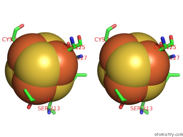
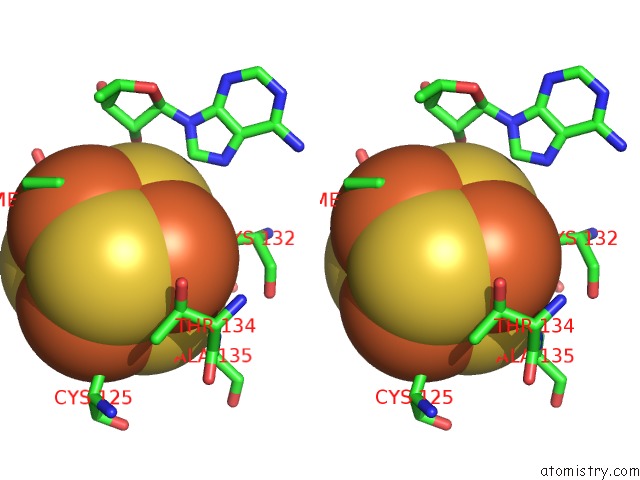
Iron binding site 4 out of 8 in 5hr6
Go back to
Iron binding site 4 out
of 8 in the X-Ray Crystal Structure of C118A Rlmn with Cross-Linked Trna Purified From Escherichia Coli
Mono view
Stereo pair view
Mono view
Stereo pair view
A full contact list of Iron with other atoms in the Fe binding
site number 4 of X-Ray Crystal Structure of C118A Rlmn with Cross-Linked Trna Purified From Escherichia Coli within 5.0Å range:
|
Iron binding site 5 out of 8 in 5hr6
Go back to
Iron binding site 5 out
of 8 in the X-Ray Crystal Structure of C118A Rlmn with Cross-Linked Trna Purified From Escherichia Coli
Mono view
Stereo pair view
Mono view
Stereo pair view
A full contact list of Iron with other atoms in the Fe binding
site number 5 of X-Ray Crystal Structure of C118A Rlmn with Cross-Linked Trna Purified From Escherichia Coli within 5.0Å range:
|
Iron binding site 6 out of 8 in 5hr6
Go back to
Iron binding site 6 out
of 8 in the X-Ray Crystal Structure of C118A Rlmn with Cross-Linked Trna Purified From Escherichia Coli
Mono view
Stereo pair view
Mono view
Stereo pair view
A full contact list of Iron with other atoms in the Fe binding
site number 6 of X-Ray Crystal Structure of C118A Rlmn with Cross-Linked Trna Purified From Escherichia Coli within 5.0Å range:
|
Iron binding site 7 out of 8 in 5hr6
Go back to
Iron binding site 7 out
of 8 in the X-Ray Crystal Structure of C118A Rlmn with Cross-Linked Trna Purified From Escherichia Coli
Mono view
Stereo pair view
Mono view
Stereo pair view
A full contact list of Iron with other atoms in the Fe binding
site number 7 of X-Ray Crystal Structure of C118A Rlmn with Cross-Linked Trna Purified From Escherichia Coli within 5.0Å range:
|
Iron binding site 8 out of 8 in 5hr6
Go back to
Iron binding site 8 out
of 8 in the X-Ray Crystal Structure of C118A Rlmn with Cross-Linked Trna Purified From Escherichia Coli
Mono view
Stereo pair view
Mono view
Stereo pair view
A full contact list of Iron with other atoms in the Fe binding
site number 8 of X-Ray Crystal Structure of C118A Rlmn with Cross-Linked Trna Purified From Escherichia Coli within 5.0Å range:
|
Reference:
E.L.Schwalm,
T.L.Grove,
S.J.Booker,
A.K.Boal.
Crystallographic Capture of A Radical S-Adenosylmethionine Enzyme in the Act of Modifying Trna. Science V. 352 309 2016.
ISSN: ESSN 1095-9203
PubMed: 27081063
DOI: 10.1126/SCIENCE.AAD5367
Page generated: Tue Aug 6 01:58:42 2024
ISSN: ESSN 1095-9203
PubMed: 27081063
DOI: 10.1126/SCIENCE.AAD5367
Last articles
Zn in 9JYWZn in 9IR4
Zn in 9IR3
Zn in 9GMX
Zn in 9GMW
Zn in 9JEJ
Zn in 9ERF
Zn in 9ERE
Zn in 9EGV
Zn in 9EGW