Iron »
PDB 5j85-5js5 »
5jom »
Iron in PDB 5jom: X-Ray Structure of Co-Bound Sperm Whale Myoglobin Using A Fixed Target Crystallography Chip
Protein crystallography data
The structure of X-Ray Structure of Co-Bound Sperm Whale Myoglobin Using A Fixed Target Crystallography Chip, PDB code: 5jom
was solved by
S.Oghbaey,
A.Sarracini,
H.M.Ginn,
O.Pare-Labrosse,
A.Kuo,
A.Marx,
S.W.Epp,
D.A.Sherrell,
B.T.Eger,
Y.Zhong,
R.Loch,
V.Mariani,
R.Alonso-Mori,
S.Nelson,
H.T.Lemke,
R.L.Owen,
A.R.Pearson,
D.I.Stuart,
O.P.Ernst,
H.M.Mueller-Werkmeister,
R.J.D.Miller,
with X-Ray Crystallography technique. A brief refinement statistics is given in the table below:
| Resolution Low / High (Å) | 33.43 / 1.90 |
| Space group | P 21 21 21 |
| Cell size a, b, c (Å), α, β, γ (°) | 38.130, 46.483, 84.521, 90.00, 90.00, 90.00 |
| R / Rfree (%) | 21.2 / 25.9 |
Iron Binding Sites:
The binding sites of Iron atom in the X-Ray Structure of Co-Bound Sperm Whale Myoglobin Using A Fixed Target Crystallography Chip
(pdb code 5jom). This binding sites where shown within
5.0 Angstroms radius around Iron atom.
In total only one binding site of Iron was determined in the X-Ray Structure of Co-Bound Sperm Whale Myoglobin Using A Fixed Target Crystallography Chip, PDB code: 5jom:
In total only one binding site of Iron was determined in the X-Ray Structure of Co-Bound Sperm Whale Myoglobin Using A Fixed Target Crystallography Chip, PDB code: 5jom:
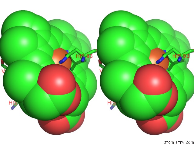
Iron binding site 1 out of 1 in 5jom
Go back to
Iron binding site 1 out
of 1 in the X-Ray Structure of Co-Bound Sperm Whale Myoglobin Using A Fixed Target Crystallography Chip
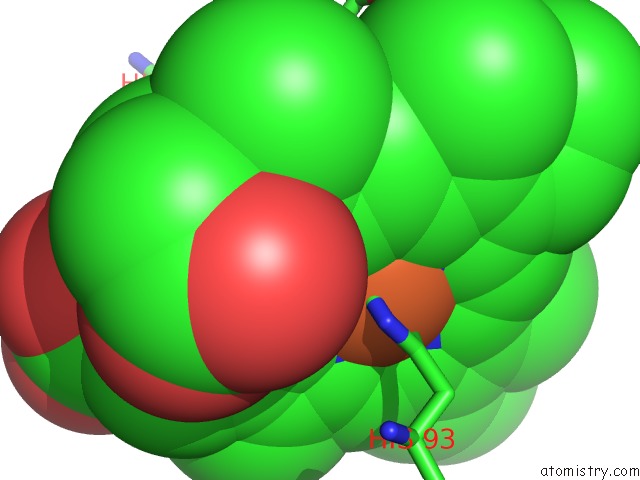
Mono view
Stereo pair view
Mono view
Stereo pair view
A full contact list of Iron with other atoms in the Fe binding
site number 1 of X-Ray Structure of Co-Bound Sperm Whale Myoglobin Using A Fixed Target Crystallography Chip within 5.0Å range:
|
Reference:
S.Oghbaey,
A.Sarracini,
H.M.Ginn,
O.Pare-Labrosse,
A.Kuo,
A.Marx,
S.W.Epp,
D.A.Sherrell,
B.T.Eger,
Y.Zhong,
R.Loch,
V.Mariani,
R.Alonso-Mori,
S.Nelson,
H.T.Lemke,
R.L.Owen,
A.R.Pearson,
D.I.Stuart,
O.P.Ernst,
H.M.Mueller-Werkmeister,
R.J.Miller.
Fixed Target Combined with Spectral Mapping: Approaching 100% Hit Rates For Serial Crystallography. Acta Crystallogr D Struct V. 72 944 2016BIOL.
ISSN: ISSN 2059-7983
PubMed: 27487825
DOI: 10.1107/S2059798316010834
Page generated: Tue Aug 6 02:34:34 2024
ISSN: ISSN 2059-7983
PubMed: 27487825
DOI: 10.1107/S2059798316010834
Last articles
Zn in 9J0NZn in 9J0O
Zn in 9J0P
Zn in 9FJX
Zn in 9EKB
Zn in 9C0F
Zn in 9CAH
Zn in 9CH0
Zn in 9CH3
Zn in 9CH1