Iron »
PDB 5jsh-5kja »
5jtd »
Iron in PDB 5jtd: Crystal Structure of the Ru(Bpy)2PHENA Functionalized P450 BM3 L407C Heme Domain Mutant in Complex with Dmso.
Enzymatic activity of Crystal Structure of the Ru(Bpy)2PHENA Functionalized P450 BM3 L407C Heme Domain Mutant in Complex with Dmso.
All present enzymatic activity of Crystal Structure of the Ru(Bpy)2PHENA Functionalized P450 BM3 L407C Heme Domain Mutant in Complex with Dmso.:
1.14.14.1; 1.6.2.4;
1.14.14.1; 1.6.2.4;
Protein crystallography data
The structure of Crystal Structure of the Ru(Bpy)2PHENA Functionalized P450 BM3 L407C Heme Domain Mutant in Complex with Dmso., PDB code: 5jtd
was solved by
M.Kloos,
with X-Ray Crystallography technique. A brief refinement statistics is given in the table below:
| Resolution Low / High (Å) | 47.36 / 1.50 |
| Space group | P 1 21 1 |
| Cell size a, b, c (Å), α, β, γ (°) | 58.690, 145.390, 62.900, 90.00, 97.08, 90.00 |
| R / Rfree (%) | 17.3 / 19.8 |
Other elements in 5jtd:
The structure of Crystal Structure of the Ru(Bpy)2PHENA Functionalized P450 BM3 L407C Heme Domain Mutant in Complex with Dmso. also contains other interesting chemical elements:
| Ruthenium | (Ru) | 2 atoms |
Iron Binding Sites:
The binding sites of Iron atom in the Crystal Structure of the Ru(Bpy)2PHENA Functionalized P450 BM3 L407C Heme Domain Mutant in Complex with Dmso.
(pdb code 5jtd). This binding sites where shown within
5.0 Angstroms radius around Iron atom.
In total 2 binding sites of Iron where determined in the Crystal Structure of the Ru(Bpy)2PHENA Functionalized P450 BM3 L407C Heme Domain Mutant in Complex with Dmso., PDB code: 5jtd:
Jump to Iron binding site number: 1; 2;
In total 2 binding sites of Iron where determined in the Crystal Structure of the Ru(Bpy)2PHENA Functionalized P450 BM3 L407C Heme Domain Mutant in Complex with Dmso., PDB code: 5jtd:
Jump to Iron binding site number: 1; 2;
Iron binding site 1 out of 2 in 5jtd
Go back to
Iron binding site 1 out
of 2 in the Crystal Structure of the Ru(Bpy)2PHENA Functionalized P450 BM3 L407C Heme Domain Mutant in Complex with Dmso.
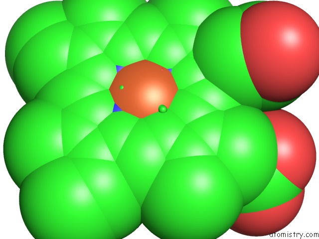
Mono view
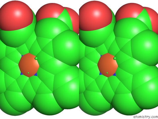
Stereo pair view
Mono view
Stereo pair view
A full contact list of Iron with other atoms in the Fe binding
site number 1 of Crystal Structure of the Ru(Bpy)2PHENA Functionalized P450 BM3 L407C Heme Domain Mutant in Complex with Dmso. within 5.0Å range:
|
Iron binding site 2 out of 2 in 5jtd
Go back to
Iron binding site 2 out
of 2 in the Crystal Structure of the Ru(Bpy)2PHENA Functionalized P450 BM3 L407C Heme Domain Mutant in Complex with Dmso.
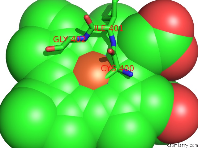
Mono view
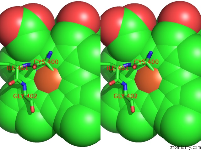
Stereo pair view
Mono view
Stereo pair view
A full contact list of Iron with other atoms in the Fe binding
site number 2 of Crystal Structure of the Ru(Bpy)2PHENA Functionalized P450 BM3 L407C Heme Domain Mutant in Complex with Dmso. within 5.0Å range:
|
Reference:
J.Spradlin,
D.Lee,
S.Mahadevan,
M.Mahomed,
L.Tang,
Q.Lam,
A.Colbert,
O.S.Shafaat,
D.Goodin,
M.Kloos,
M.Kato,
L.E.Cheruzel.
Insights Into An Efficient Light-Driven Hybrid P450 BM3 Enzyme From Crystallographic, Spectroscopic and Biochemical Studies. Biochim.Biophys.Acta V.1864 1732 2016.
ISSN: ISSN 0006-3002
PubMed: 27639964
DOI: 10.1016/J.BBAPAP.2016.09.005
Page generated: Tue Aug 6 02:56:22 2024
ISSN: ISSN 0006-3002
PubMed: 27639964
DOI: 10.1016/J.BBAPAP.2016.09.005
Last articles
Zn in 9J0NZn in 9J0O
Zn in 9J0P
Zn in 9FJX
Zn in 9EKB
Zn in 9C0F
Zn in 9CAH
Zn in 9CH0
Zn in 9CH3
Zn in 9CH1