Iron »
PDB 5jsh-5kja »
5k53 »
Iron in PDB 5k53: Crystal Structures of Aldehyde Deformylating Oxygenase From Oscillatoria Sp. KNUA011
Protein crystallography data
The structure of Crystal Structures of Aldehyde Deformylating Oxygenase From Oscillatoria Sp. KNUA011, PDB code: 5k53
was solved by
A.K.Park,
H-.W.Kim,
with X-Ray Crystallography technique. A brief refinement statistics is given in the table below:
| Resolution Low / High (Å) | 50.01 / 1.80 |
| Space group | P 21 21 21 |
| Cell size a, b, c (Å), α, β, γ (°) | 55.012, 78.265, 108.861, 90.00, 90.00, 90.00 |
| R / Rfree (%) | 19.3 / 23.4 |
Iron Binding Sites:
The binding sites of Iron atom in the Crystal Structures of Aldehyde Deformylating Oxygenase From Oscillatoria Sp. KNUA011
(pdb code 5k53). This binding sites where shown within
5.0 Angstroms radius around Iron atom.
In total 4 binding sites of Iron where determined in the Crystal Structures of Aldehyde Deformylating Oxygenase From Oscillatoria Sp. KNUA011, PDB code: 5k53:
Jump to Iron binding site number: 1; 2; 3; 4;
In total 4 binding sites of Iron where determined in the Crystal Structures of Aldehyde Deformylating Oxygenase From Oscillatoria Sp. KNUA011, PDB code: 5k53:
Jump to Iron binding site number: 1; 2; 3; 4;
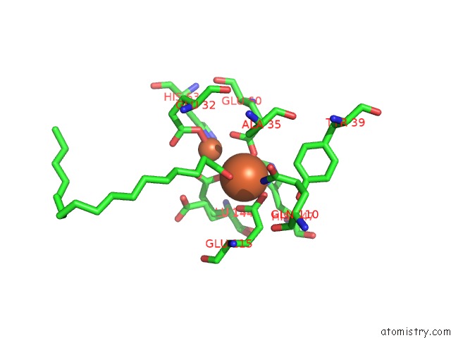
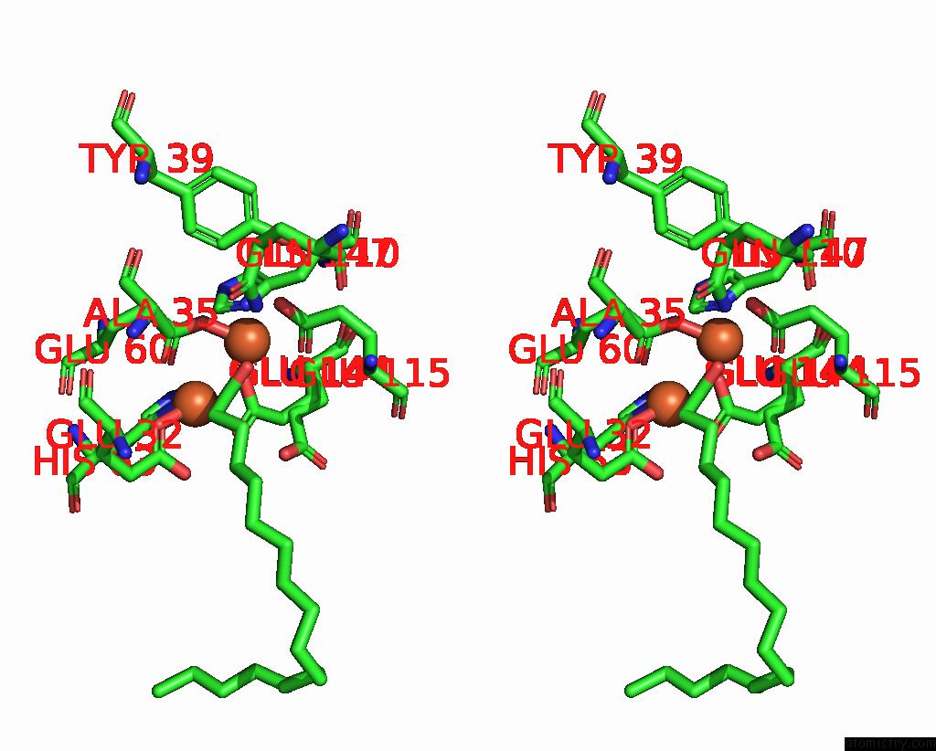
Iron binding site 1 out of 4 in 5k53
Go back to
Iron binding site 1 out
of 4 in the Crystal Structures of Aldehyde Deformylating Oxygenase From Oscillatoria Sp. KNUA011
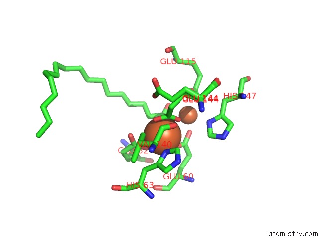
Mono view
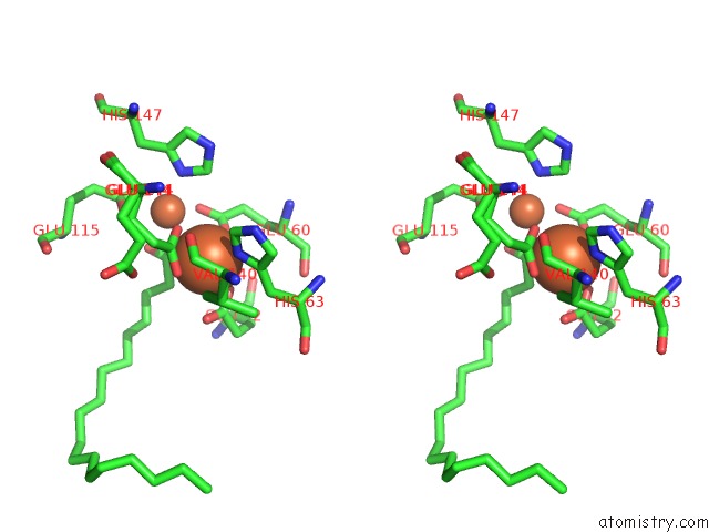
Stereo pair view
Mono view
Stereo pair view
A full contact list of Iron with other atoms in the Fe binding
site number 1 of Crystal Structures of Aldehyde Deformylating Oxygenase From Oscillatoria Sp. KNUA011 within 5.0Å range:
|
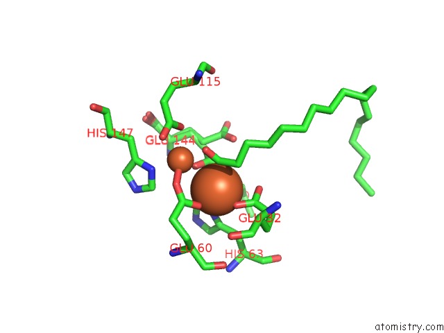
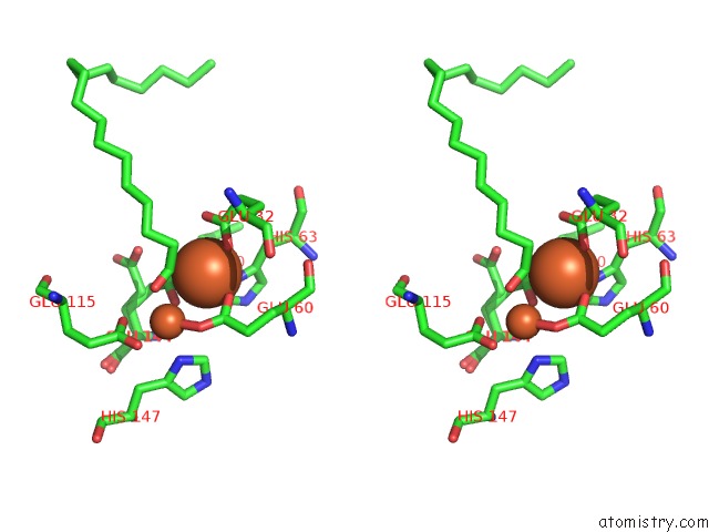
Iron binding site 2 out of 4 in 5k53
Go back to
Iron binding site 2 out
of 4 in the Crystal Structures of Aldehyde Deformylating Oxygenase From Oscillatoria Sp. KNUA011
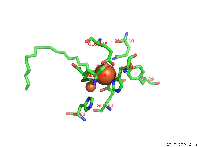
Mono view
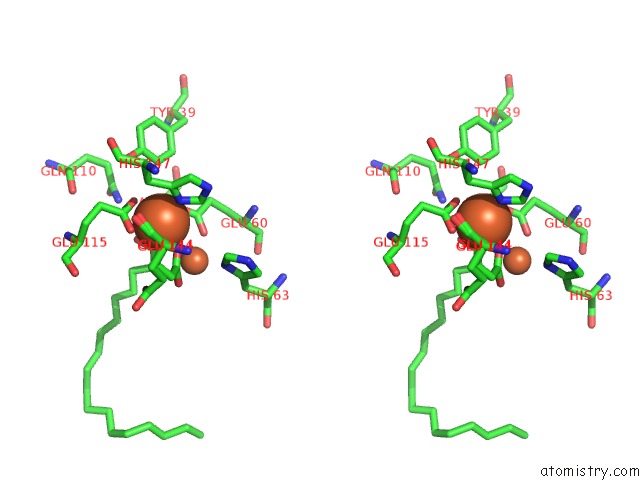
Stereo pair view
Mono view
Stereo pair view
A full contact list of Iron with other atoms in the Fe binding
site number 2 of Crystal Structures of Aldehyde Deformylating Oxygenase From Oscillatoria Sp. KNUA011 within 5.0Å range:
|
Iron binding site 3 out of 4 in 5k53
Go back to
Iron binding site 3 out
of 4 in the Crystal Structures of Aldehyde Deformylating Oxygenase From Oscillatoria Sp. KNUA011
Mono view
Stereo pair view
Mono view
Stereo pair view
A full contact list of Iron with other atoms in the Fe binding
site number 3 of Crystal Structures of Aldehyde Deformylating Oxygenase From Oscillatoria Sp. KNUA011 within 5.0Å range:
|
Iron binding site 4 out of 4 in 5k53
Go back to
Iron binding site 4 out
of 4 in the Crystal Structures of Aldehyde Deformylating Oxygenase From Oscillatoria Sp. KNUA011
Mono view
Stereo pair view
Mono view
Stereo pair view
A full contact list of Iron with other atoms in the Fe binding
site number 4 of Crystal Structures of Aldehyde Deformylating Oxygenase From Oscillatoria Sp. KNUA011 within 5.0Å range:
|
Reference:
A.K.Park,
I.S.Kim,
B.W.Jeon,
S.J.Roh,
M.Y.Ryu,
H.R.Baek,
S.W.Jo,
Y.S.Kim,
H.Park,
J.H.Lee,
H.S.Yoon,
H.W.Kim.
Crystal Structures of Aldehyde Deformylating Oxygenase From Limnothrix Sp. KNUA012 and Oscillatoria Sp. KNUA011. Biochem.Biophys.Res.Commun. V. 477 395 2016.
ISSN: ESSN 1090-2104
PubMed: 27329814
DOI: 10.1016/J.BBRC.2016.06.090
Page generated: Tue Aug 6 03:00:44 2024
ISSN: ESSN 1090-2104
PubMed: 27329814
DOI: 10.1016/J.BBRC.2016.06.090
Last articles
Fe in 2YXOFe in 2YRS
Fe in 2YXC
Fe in 2YNM
Fe in 2YVJ
Fe in 2YP1
Fe in 2YU2
Fe in 2YU1
Fe in 2YQB
Fe in 2YOO