Iron »
PDB 5kjb-5ktr »
5kkk »
Iron in PDB 5kkk: 1.7-Angstrom in Situ Mylar Structure of Sperm Whale Myoglobin (Swmb- Co) at 100 K
Protein crystallography data
The structure of 1.7-Angstrom in Situ Mylar Structure of Sperm Whale Myoglobin (Swmb- Co) at 100 K, PDB code: 5kkk
was solved by
J.Broecker,
O.P.Ernst,
with X-Ray Crystallography technique. A brief refinement statistics is given in the table below:
| Resolution Low / High (Å) | 24.91 / 1.70 |
| Space group | P 6 |
| Cell size a, b, c (Å), α, β, γ (°) | 90.770, 90.770, 45.671, 90.00, 90.00, 120.00 |
| R / Rfree (%) | 17.6 / 21.5 |
Other elements in 5kkk:
The structure of 1.7-Angstrom in Situ Mylar Structure of Sperm Whale Myoglobin (Swmb- Co) at 100 K also contains other interesting chemical elements:
| Chlorine | (Cl) | 2 atoms |
Iron Binding Sites:
The binding sites of Iron atom in the 1.7-Angstrom in Situ Mylar Structure of Sperm Whale Myoglobin (Swmb- Co) at 100 K
(pdb code 5kkk). This binding sites where shown within
5.0 Angstroms radius around Iron atom.
In total only one binding site of Iron was determined in the 1.7-Angstrom in Situ Mylar Structure of Sperm Whale Myoglobin (Swmb- Co) at 100 K, PDB code: 5kkk:
In total only one binding site of Iron was determined in the 1.7-Angstrom in Situ Mylar Structure of Sperm Whale Myoglobin (Swmb- Co) at 100 K, PDB code: 5kkk:
Iron binding site 1 out of 1 in 5kkk
Go back to
Iron binding site 1 out
of 1 in the 1.7-Angstrom in Situ Mylar Structure of Sperm Whale Myoglobin (Swmb- Co) at 100 K
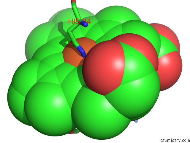
Mono view
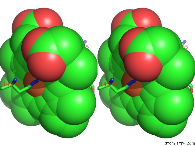
Stereo pair view
Mono view
Stereo pair view
A full contact list of Iron with other atoms in the Fe binding
site number 1 of 1.7-Angstrom in Situ Mylar Structure of Sperm Whale Myoglobin (Swmb- Co) at 100 K within 5.0Å range:
|
Reference:
J.Broecker,
V.Klingel,
W.L.Ou,
A.R.Balo,
D.J.Kissick,
C.M.Ogata,
A.Kuo,
O.P.Ernst.
A Versatile System For High-Throughput in Situ X-Ray Screening and Data Collection of Soluble and Membrane-Protein Crystals. Cryst Growth Des V. 16 6318 2016.
ISSN: ISSN 1528-7483
PubMed: 28261000
DOI: 10.1021/ACS.CGD.6B00950
Page generated: Tue Aug 5 22:37:37 2025
ISSN: ISSN 1528-7483
PubMed: 28261000
DOI: 10.1021/ACS.CGD.6B00950
Last articles
Fe in 5OBAFe in 5O31
Fe in 5OD5
Fe in 5OCF
Fe in 5OBP
Fe in 5OCB
Fe in 5OC0
Fe in 5OBI
Fe in 5OBO
Fe in 5OAY