Iron »
PDB 5kjb-5ktr »
5ksf »
Iron in PDB 5ksf: Crystal Structure of the D141A Variant of the Catalase-Peroxidase From B. Pseudomallei Treated with Acetate
Enzymatic activity of Crystal Structure of the D141A Variant of the Catalase-Peroxidase From B. Pseudomallei Treated with Acetate
All present enzymatic activity of Crystal Structure of the D141A Variant of the Catalase-Peroxidase From B. Pseudomallei Treated with Acetate:
1.11.1.21;
1.11.1.21;
Protein crystallography data
The structure of Crystal Structure of the D141A Variant of the Catalase-Peroxidase From B. Pseudomallei Treated with Acetate, PDB code: 5ksf
was solved by
P.C.Loewen,
with X-Ray Crystallography technique. A brief refinement statistics is given in the table below:
| Resolution Low / High (Å) | 48.38 / 1.75 |
| Space group | P 21 21 21 |
| Cell size a, b, c (Å), α, β, γ (°) | 100.692, 115.474, 174.586, 90.00, 90.00, 90.00 |
| R / Rfree (%) | 14.8 / 17.5 |
Other elements in 5ksf:
The structure of Crystal Structure of the D141A Variant of the Catalase-Peroxidase From B. Pseudomallei Treated with Acetate also contains other interesting chemical elements:
| Chlorine | (Cl) | 2 atoms |
| Sodium | (Na) | 2 atoms |
Iron Binding Sites:
The binding sites of Iron atom in the Crystal Structure of the D141A Variant of the Catalase-Peroxidase From B. Pseudomallei Treated with Acetate
(pdb code 5ksf). This binding sites where shown within
5.0 Angstroms radius around Iron atom.
In total 2 binding sites of Iron where determined in the Crystal Structure of the D141A Variant of the Catalase-Peroxidase From B. Pseudomallei Treated with Acetate, PDB code: 5ksf:
Jump to Iron binding site number: 1; 2;
In total 2 binding sites of Iron where determined in the Crystal Structure of the D141A Variant of the Catalase-Peroxidase From B. Pseudomallei Treated with Acetate, PDB code: 5ksf:
Jump to Iron binding site number: 1; 2;
Iron binding site 1 out of 2 in 5ksf
Go back to
Iron binding site 1 out
of 2 in the Crystal Structure of the D141A Variant of the Catalase-Peroxidase From B. Pseudomallei Treated with Acetate
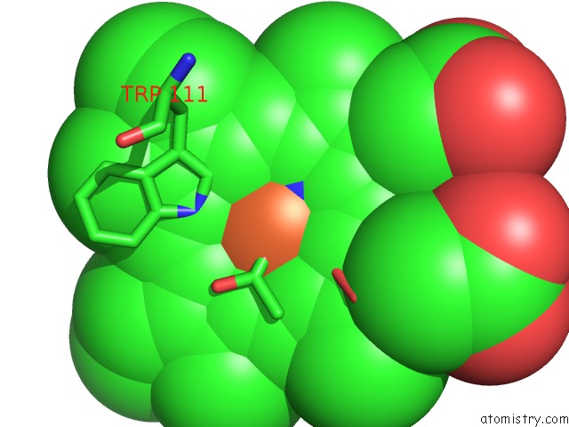
Mono view
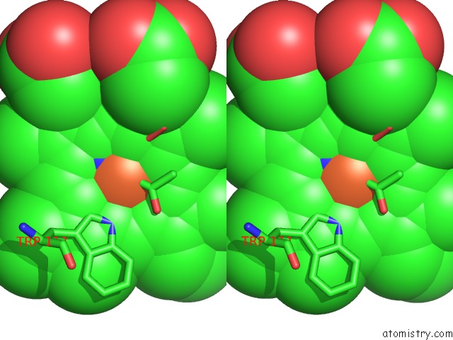
Stereo pair view
Mono view
Stereo pair view
A full contact list of Iron with other atoms in the Fe binding
site number 1 of Crystal Structure of the D141A Variant of the Catalase-Peroxidase From B. Pseudomallei Treated with Acetate within 5.0Å range:
|
Iron binding site 2 out of 2 in 5ksf
Go back to
Iron binding site 2 out
of 2 in the Crystal Structure of the D141A Variant of the Catalase-Peroxidase From B. Pseudomallei Treated with Acetate
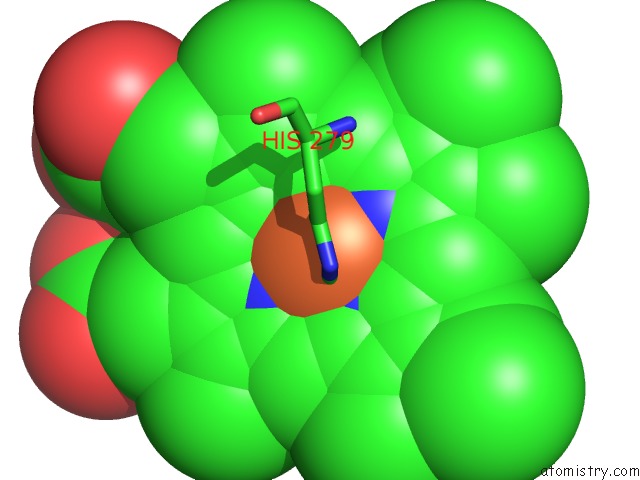
Mono view
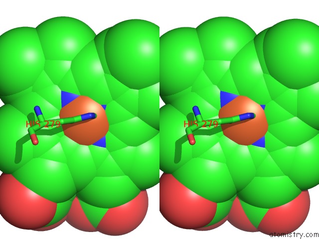
Stereo pair view
Mono view
Stereo pair view
A full contact list of Iron with other atoms in the Fe binding
site number 2 of Crystal Structure of the D141A Variant of the Catalase-Peroxidase From B. Pseudomallei Treated with Acetate within 5.0Å range:
|
Reference:
M.Machuqueiro,
B.Victor,
J.Switala,
J.Villanueva,
C.Rovira,
I.Fita,
P.C.Loewen.
The Catalase Activity of Catalase-Peroxidases Is Modulated By Changes in the Pka of the Distal Histidine. Biochemistry V. 56 2271 2017.
ISSN: ISSN 1520-4995
PubMed: 28409923
DOI: 10.1021/ACS.BIOCHEM.6B01276
Page generated: Tue Aug 6 03:38:32 2024
ISSN: ISSN 1520-4995
PubMed: 28409923
DOI: 10.1021/ACS.BIOCHEM.6B01276
Last articles
Zn in 9J0NZn in 9J0O
Zn in 9J0P
Zn in 9FJX
Zn in 9EKB
Zn in 9C0F
Zn in 9CAH
Zn in 9CH0
Zn in 9CH3
Zn in 9CH1