Iron »
PDB 5kts-5lg8 »
5lbh »
Iron in PDB 5lbh: Crystal Structure of Helicobacter Cinaedi Caip
Protein crystallography data
The structure of Crystal Structure of Helicobacter Cinaedi Caip, PDB code: 5lbh
was solved by
G.Zanotti,
F.Valesse,
G.Codolo,
M.De Bernard,
with X-Ray Crystallography technique. A brief refinement statistics is given in the table below:
| Resolution Low / High (Å) | 46.53 / 2.55 |
| Space group | P 1 21 1 |
| Cell size a, b, c (Å), α, β, γ (°) | 89.746, 135.918, 93.445, 90.00, 114.50, 90.00 |
| R / Rfree (%) | 18.7 / 24.8 |
Iron Binding Sites:
Pages:
>>> Page 1 <<< Page 2, Binding sites: 11 - 12;Binding sites:
The binding sites of Iron atom in the Crystal Structure of Helicobacter Cinaedi Caip (pdb code 5lbh). This binding sites where shown within 5.0 Angstroms radius around Iron atom.In total 12 binding sites of Iron where determined in the Crystal Structure of Helicobacter Cinaedi Caip, PDB code: 5lbh:
Jump to Iron binding site number: 1; 2; 3; 4; 5; 6; 7; 8; 9; 10;
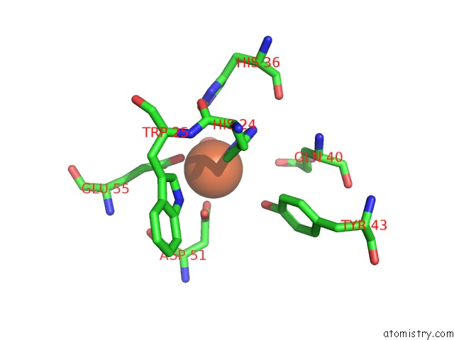
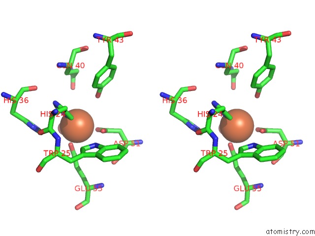
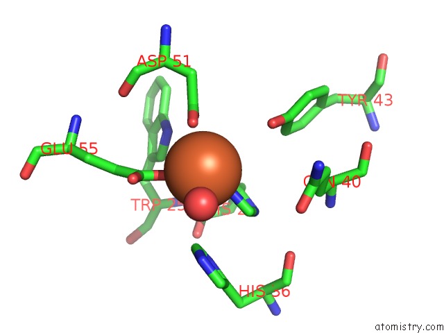
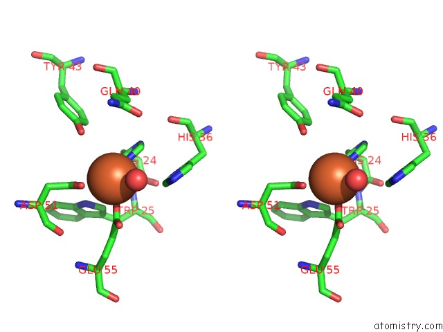
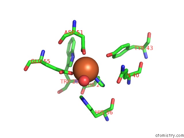
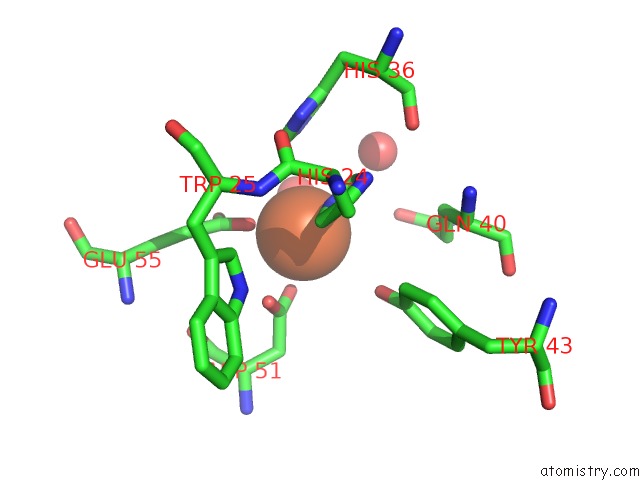
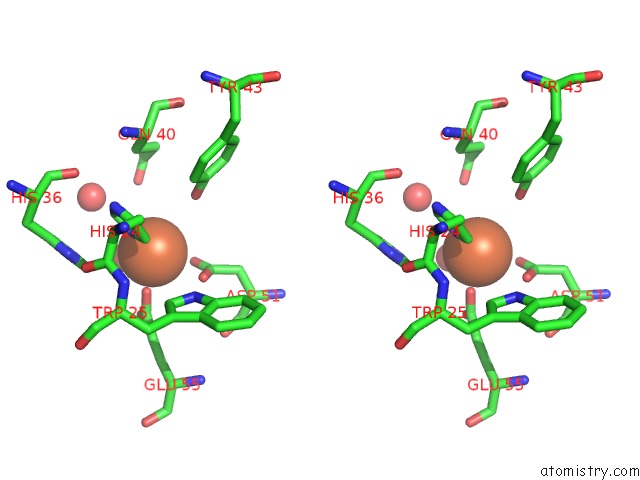
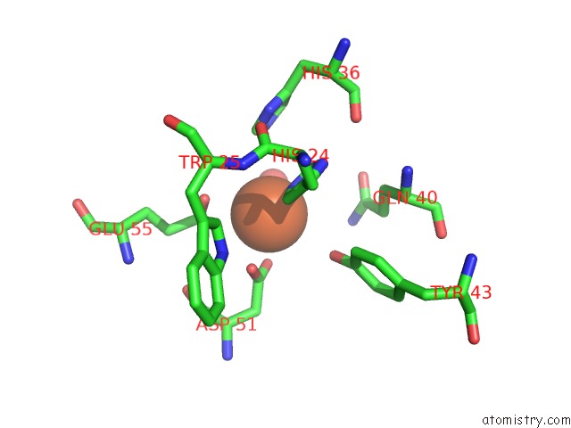
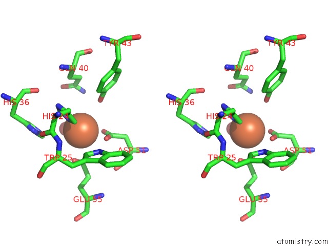
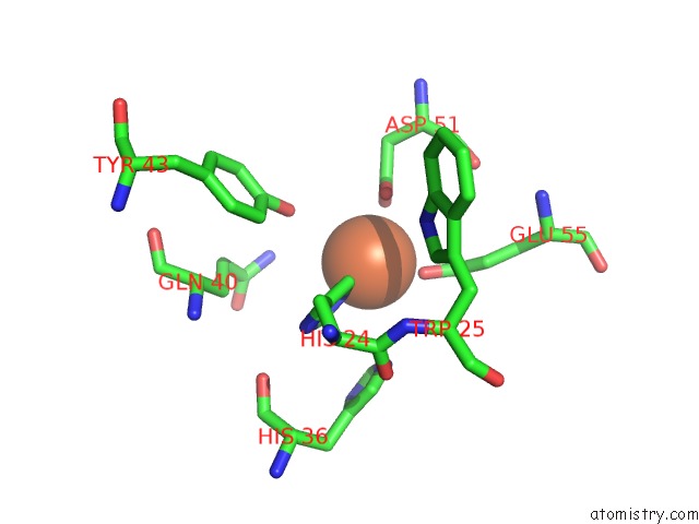
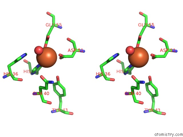
Iron binding site 1 out of 12 in 5lbh
Go back to
Iron binding site 1 out
of 12 in the Crystal Structure of Helicobacter Cinaedi Caip
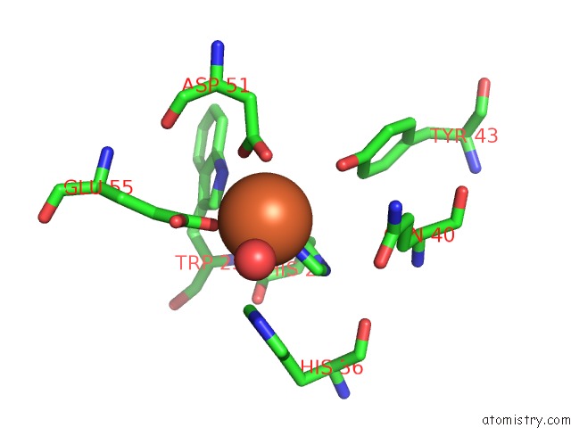
Mono view
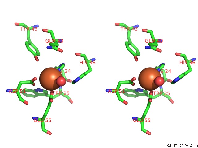
Stereo pair view
Mono view
Stereo pair view
A full contact list of Iron with other atoms in the Fe binding
site number 1 of Crystal Structure of Helicobacter Cinaedi Caip within 5.0Å range:
|
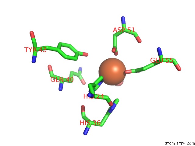
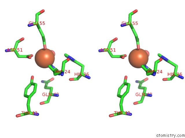
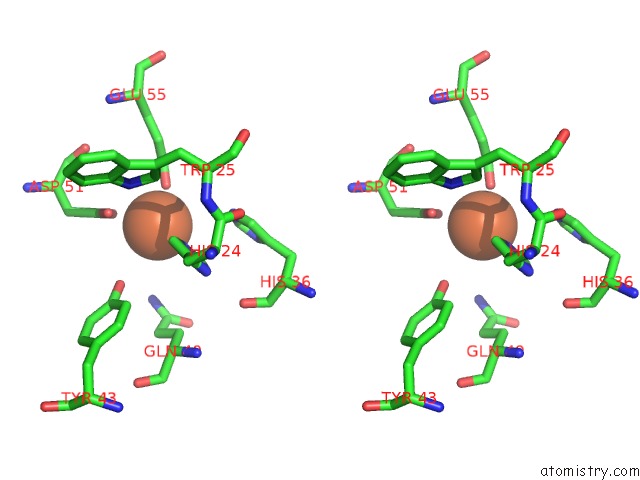
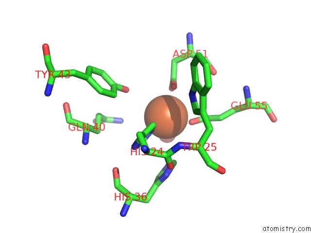
Iron binding site 2 out of 12 in 5lbh
Go back to
Iron binding site 2 out
of 12 in the Crystal Structure of Helicobacter Cinaedi Caip
Mono view
Stereo pair view
Mono view
Stereo pair view
A full contact list of Iron with other atoms in the Fe binding
site number 2 of Crystal Structure of Helicobacter Cinaedi Caip within 5.0Å range:
|
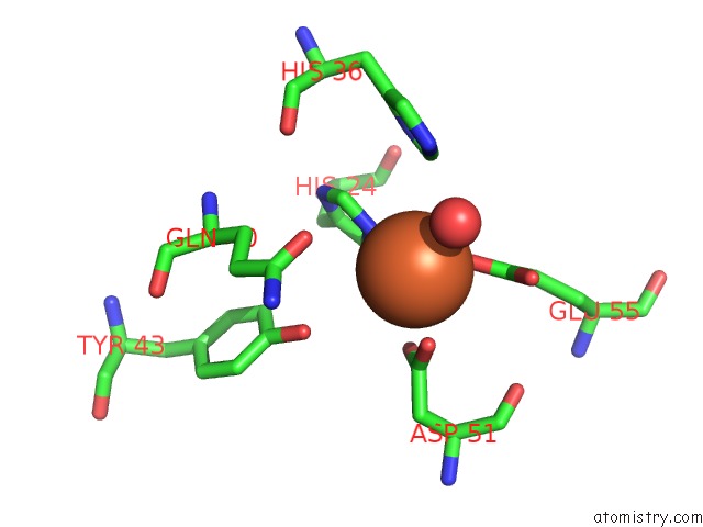
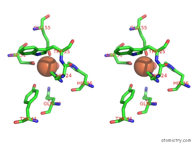
Iron binding site 3 out of 12 in 5lbh
Go back to
Iron binding site 3 out
of 12 in the Crystal Structure of Helicobacter Cinaedi Caip
Mono view
Stereo pair view
Mono view
Stereo pair view
A full contact list of Iron with other atoms in the Fe binding
site number 3 of Crystal Structure of Helicobacter Cinaedi Caip within 5.0Å range:
|
Iron binding site 4 out of 12 in 5lbh
Go back to
Iron binding site 4 out
of 12 in the Crystal Structure of Helicobacter Cinaedi Caip
Mono view
Stereo pair view
Mono view
Stereo pair view
A full contact list of Iron with other atoms in the Fe binding
site number 4 of Crystal Structure of Helicobacter Cinaedi Caip within 5.0Å range:
|
Iron binding site 5 out of 12 in 5lbh
Go back to
Iron binding site 5 out
of 12 in the Crystal Structure of Helicobacter Cinaedi Caip
Mono view

Stereo pair view
Mono view
Stereo pair view
A full contact list of Iron with other atoms in the Fe binding
site number 5 of Crystal Structure of Helicobacter Cinaedi Caip within 5.0Å range:
|
Iron binding site 6 out of 12 in 5lbh
Go back to
Iron binding site 6 out
of 12 in the Crystal Structure of Helicobacter Cinaedi Caip
Mono view
Stereo pair view
Mono view
Stereo pair view
A full contact list of Iron with other atoms in the Fe binding
site number 6 of Crystal Structure of Helicobacter Cinaedi Caip within 5.0Å range:
|
Iron binding site 7 out of 12 in 5lbh
Go back to
Iron binding site 7 out
of 12 in the Crystal Structure of Helicobacter Cinaedi Caip
Mono view
Stereo pair view
Mono view
Stereo pair view
A full contact list of Iron with other atoms in the Fe binding
site number 7 of Crystal Structure of Helicobacter Cinaedi Caip within 5.0Å range:
|
Iron binding site 8 out of 12 in 5lbh
Go back to
Iron binding site 8 out
of 12 in the Crystal Structure of Helicobacter Cinaedi Caip
Mono view
Stereo pair view
Mono view
Stereo pair view
A full contact list of Iron with other atoms in the Fe binding
site number 8 of Crystal Structure of Helicobacter Cinaedi Caip within 5.0Å range:
|
Iron binding site 9 out of 12 in 5lbh
Go back to
Iron binding site 9 out
of 12 in the Crystal Structure of Helicobacter Cinaedi Caip
Mono view
Stereo pair view
Mono view
Stereo pair view
A full contact list of Iron with other atoms in the Fe binding
site number 9 of Crystal Structure of Helicobacter Cinaedi Caip within 5.0Å range:
|
Iron binding site 10 out of 12 in 5lbh
Go back to
Iron binding site 10 out
of 12 in the Crystal Structure of Helicobacter Cinaedi Caip
Mono view
Stereo pair view
Mono view
Stereo pair view
A full contact list of Iron with other atoms in the Fe binding
site number 10 of Crystal Structure of Helicobacter Cinaedi Caip within 5.0Å range:
|
Reference:
M.M.D'elios,
F.Vallese,
N.Capitani,
M.Benagiano,
M.L.Bernardini,
M.Rossi,
G.P.Rossi,
M.Ferrari,
C.T.Baldari,
G.Zanotti,
M.De Bernard,
G.Codolo.
The Helicobacter Cinaedi Antigen Caip Participates in Atherosclerotic Inflammation By Promoting the Differentiation of Macrophages in Foam Cells. Sci Rep V. 7 40515 2017.
ISSN: ESSN 2045-2322
PubMed: 28074932
DOI: 10.1038/SREP40515
Page generated: Tue Aug 6 04:09:22 2024
ISSN: ESSN 2045-2322
PubMed: 28074932
DOI: 10.1038/SREP40515
Last articles
Zn in 9MJ5Zn in 9HNW
Zn in 9G0L
Zn in 9FNE
Zn in 9DZN
Zn in 9E0I
Zn in 9D32
Zn in 9DAK
Zn in 8ZXC
Zn in 8ZUF