Iron »
PDB 5kts-5lg8 »
5lfa »
Iron in PDB 5lfa: Crystal Structure of Iron-Sulfur Cluster Containing Bacterial (6-4) Photolyase Phrb - Y424F Mutant with Impaired Dna Repair Activity
Enzymatic activity of Crystal Structure of Iron-Sulfur Cluster Containing Bacterial (6-4) Photolyase Phrb - Y424F Mutant with Impaired Dna Repair Activity
All present enzymatic activity of Crystal Structure of Iron-Sulfur Cluster Containing Bacterial (6-4) Photolyase Phrb - Y424F Mutant with Impaired Dna Repair Activity:
4.1.99.13;
4.1.99.13;
Protein crystallography data
The structure of Crystal Structure of Iron-Sulfur Cluster Containing Bacterial (6-4) Photolyase Phrb - Y424F Mutant with Impaired Dna Repair Activity, PDB code: 5lfa
was solved by
D.Kwiatkowski,
F.Zhang,
N.Krauss,
T.Lamparter,
P.Scheerer,
with X-Ray Crystallography technique. A brief refinement statistics is given in the table below:
| Resolution Low / High (Å) | 69.79 / 2.50 |
| Space group | P 2 21 21 |
| Cell size a, b, c (Å), α, β, γ (°) | 56.183, 94.441, 103.579, 90.00, 90.00, 90.00 |
| R / Rfree (%) | 18.1 / 24 |
Iron Binding Sites:
The binding sites of Iron atom in the Crystal Structure of Iron-Sulfur Cluster Containing Bacterial (6-4) Photolyase Phrb - Y424F Mutant with Impaired Dna Repair Activity
(pdb code 5lfa). This binding sites where shown within
5.0 Angstroms radius around Iron atom.
In total 4 binding sites of Iron where determined in the Crystal Structure of Iron-Sulfur Cluster Containing Bacterial (6-4) Photolyase Phrb - Y424F Mutant with Impaired Dna Repair Activity, PDB code: 5lfa:
Jump to Iron binding site number: 1; 2; 3; 4;
In total 4 binding sites of Iron where determined in the Crystal Structure of Iron-Sulfur Cluster Containing Bacterial (6-4) Photolyase Phrb - Y424F Mutant with Impaired Dna Repair Activity, PDB code: 5lfa:
Jump to Iron binding site number: 1; 2; 3; 4;
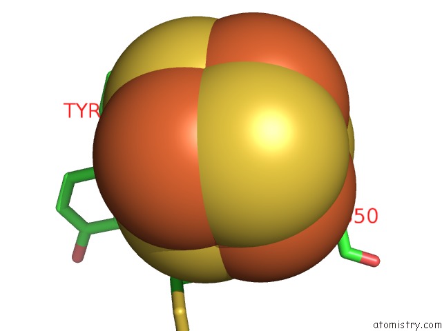
Iron binding site 1 out of 4 in 5lfa
Go back to
Iron binding site 1 out
of 4 in the Crystal Structure of Iron-Sulfur Cluster Containing Bacterial (6-4) Photolyase Phrb - Y424F Mutant with Impaired Dna Repair Activity

Mono view
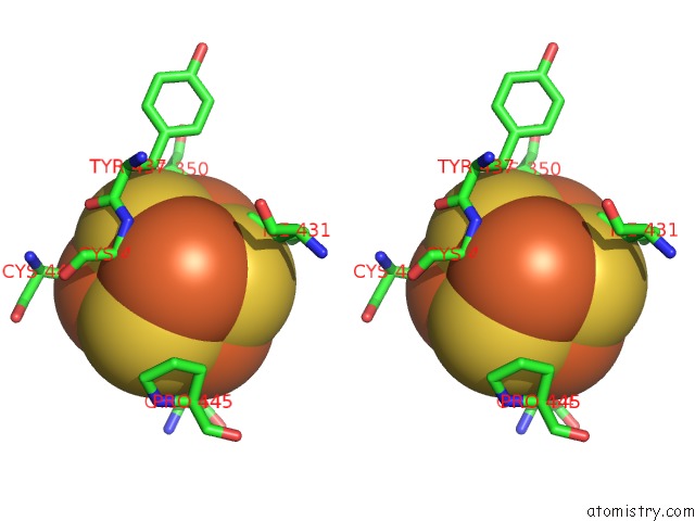
Stereo pair view
Mono view
Stereo pair view
A full contact list of Iron with other atoms in the Fe binding
site number 1 of Crystal Structure of Iron-Sulfur Cluster Containing Bacterial (6-4) Photolyase Phrb - Y424F Mutant with Impaired Dna Repair Activity within 5.0Å range:
|
Iron binding site 2 out of 4 in 5lfa
Go back to
Iron binding site 2 out
of 4 in the Crystal Structure of Iron-Sulfur Cluster Containing Bacterial (6-4) Photolyase Phrb - Y424F Mutant with Impaired Dna Repair Activity

Mono view

Stereo pair view
Mono view
Stereo pair view
A full contact list of Iron with other atoms in the Fe binding
site number 2 of Crystal Structure of Iron-Sulfur Cluster Containing Bacterial (6-4) Photolyase Phrb - Y424F Mutant with Impaired Dna Repair Activity within 5.0Å range:
|
Iron binding site 3 out of 4 in 5lfa
Go back to
Iron binding site 3 out
of 4 in the Crystal Structure of Iron-Sulfur Cluster Containing Bacterial (6-4) Photolyase Phrb - Y424F Mutant with Impaired Dna Repair Activity
Mono view

Stereo pair view
Mono view
Stereo pair view
A full contact list of Iron with other atoms in the Fe binding
site number 3 of Crystal Structure of Iron-Sulfur Cluster Containing Bacterial (6-4) Photolyase Phrb - Y424F Mutant with Impaired Dna Repair Activity within 5.0Å range:
|
Iron binding site 4 out of 4 in 5lfa
Go back to
Iron binding site 4 out
of 4 in the Crystal Structure of Iron-Sulfur Cluster Containing Bacterial (6-4) Photolyase Phrb - Y424F Mutant with Impaired Dna Repair Activity

Mono view

Stereo pair view
Mono view
Stereo pair view
A full contact list of Iron with other atoms in the Fe binding
site number 4 of Crystal Structure of Iron-Sulfur Cluster Containing Bacterial (6-4) Photolyase Phrb - Y424F Mutant with Impaired Dna Repair Activity within 5.0Å range:
|
Reference:
F.Zhang,
H.Ma,
K.Bowatte,
D.Kwiatkowski,
E.Mittmann,
H.Qasem,
N.Krau,
X.Zeng,
Z.Ren,
P.Scheerer,
X.Yang,
T.Lamparter.
Crystal Structures of Bacterial (6-4) Photolyase Mutants with Impaired Dna Repair Activity. Photochem. Photobiol. V. 93 304 2017.
ISSN: ISSN 1751-1097
PubMed: 27992645
DOI: 10.1111/PHP.12699
Page generated: Tue Aug 6 04:10:56 2024
ISSN: ISSN 1751-1097
PubMed: 27992645
DOI: 10.1111/PHP.12699
Last articles
Fe in 2YXOFe in 2YRS
Fe in 2YXC
Fe in 2YNM
Fe in 2YVJ
Fe in 2YP1
Fe in 2YU2
Fe in 2YU1
Fe in 2YQB
Fe in 2YOO