Iron »
PDB 5li6-5m2g »
5li7 »
Iron in PDB 5li7: Crystal Structure of Mycobacterium Tuberculosis CYP126A1 in Complex with 1-(3-(1H-Imidazol-1-Yl)Propyl)-3-((3S,5S,7S)-Adamantan-1-Yl)Urea
Protein crystallography data
The structure of Crystal Structure of Mycobacterium Tuberculosis CYP126A1 in Complex with 1-(3-(1H-Imidazol-1-Yl)Propyl)-3-((3S,5S,7S)-Adamantan-1-Yl)Urea, PDB code: 5li7
was solved by
C.Levy,
A.W.Munro,
D.Leys,
with X-Ray Crystallography technique. A brief refinement statistics is given in the table below:
| Resolution Low / High (Å) | 57.31 / 1.58 |
| Space group | P 21 21 21 |
| Cell size a, b, c (Å), α, β, γ (°) | 59.250, 69.910, 226.140, 90.00, 90.00, 90.00 |
| R / Rfree (%) | 17 / 20.3 |
Iron Binding Sites:
The binding sites of Iron atom in the Crystal Structure of Mycobacterium Tuberculosis CYP126A1 in Complex with 1-(3-(1H-Imidazol-1-Yl)Propyl)-3-((3S,5S,7S)-Adamantan-1-Yl)Urea
(pdb code 5li7). This binding sites where shown within
5.0 Angstroms radius around Iron atom.
In total 2 binding sites of Iron where determined in the Crystal Structure of Mycobacterium Tuberculosis CYP126A1 in Complex with 1-(3-(1H-Imidazol-1-Yl)Propyl)-3-((3S,5S,7S)-Adamantan-1-Yl)Urea, PDB code: 5li7:
Jump to Iron binding site number: 1; 2;
In total 2 binding sites of Iron where determined in the Crystal Structure of Mycobacterium Tuberculosis CYP126A1 in Complex with 1-(3-(1H-Imidazol-1-Yl)Propyl)-3-((3S,5S,7S)-Adamantan-1-Yl)Urea, PDB code: 5li7:
Jump to Iron binding site number: 1; 2;
Iron binding site 1 out of 2 in 5li7
Go back to
Iron binding site 1 out
of 2 in the Crystal Structure of Mycobacterium Tuberculosis CYP126A1 in Complex with 1-(3-(1H-Imidazol-1-Yl)Propyl)-3-((3S,5S,7S)-Adamantan-1-Yl)Urea
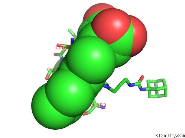
Mono view
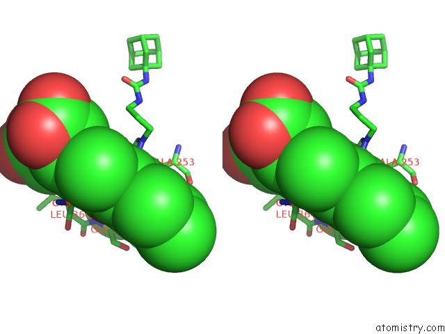
Stereo pair view
Mono view
Stereo pair view
A full contact list of Iron with other atoms in the Fe binding
site number 1 of Crystal Structure of Mycobacterium Tuberculosis CYP126A1 in Complex with 1-(3-(1H-Imidazol-1-Yl)Propyl)-3-((3S,5S,7S)-Adamantan-1-Yl)Urea within 5.0Å range:
|
Iron binding site 2 out of 2 in 5li7
Go back to
Iron binding site 2 out
of 2 in the Crystal Structure of Mycobacterium Tuberculosis CYP126A1 in Complex with 1-(3-(1H-Imidazol-1-Yl)Propyl)-3-((3S,5S,7S)-Adamantan-1-Yl)Urea
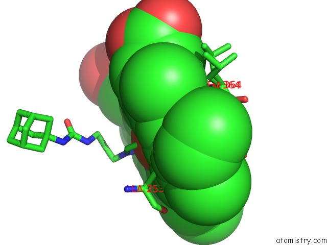
Mono view
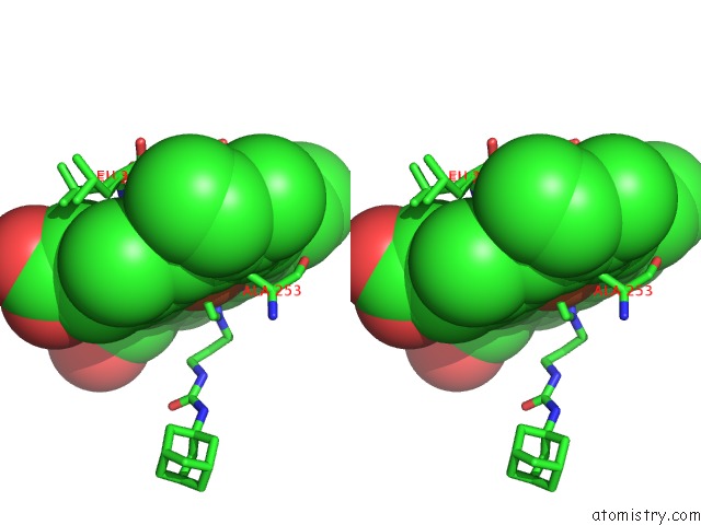
Stereo pair view
Mono view
Stereo pair view
A full contact list of Iron with other atoms in the Fe binding
site number 2 of Crystal Structure of Mycobacterium Tuberculosis CYP126A1 in Complex with 1-(3-(1H-Imidazol-1-Yl)Propyl)-3-((3S,5S,7S)-Adamantan-1-Yl)Urea within 5.0Å range:
|
Reference:
J.T.Chenge,
L.V.Duyet,
S.Swami,
K.J.Mclean,
M.E.Kavanagh,
A.G.Coyne,
S.E.Rigby,
M.R.Cheesman,
H.M.Girvan,
C.W.Levy,
B.Rupp,
J.P.Von Kries,
C.Abell,
D.Leys,
A.W.Munro.
Structural Characterization and Ligand/Inhibitor Identification Provide Functional Insights Into the Mycobacterium Tuberculosis Cytochrome P450 CYP126A1. J. Biol. Chem. V. 292 1310 2017.
ISSN: ESSN 1083-351X
PubMed: 27932461
DOI: 10.1074/JBC.M116.748822
Page generated: Tue Aug 6 04:40:12 2024
ISSN: ESSN 1083-351X
PubMed: 27932461
DOI: 10.1074/JBC.M116.748822
Last articles
Zn in 9J0NZn in 9J0O
Zn in 9J0P
Zn in 9FJX
Zn in 9EKB
Zn in 9C0F
Zn in 9CAH
Zn in 9CH0
Zn in 9CH3
Zn in 9CH1