Iron »
PDB 5li6-5m2g »
5m0t »
Iron in PDB 5m0t: Alpha-Ketoglutarate-Dependent Non-Heme Iron Oxygenase Eash
Protein crystallography data
The structure of Alpha-Ketoglutarate-Dependent Non-Heme Iron Oxygenase Eash, PDB code: 5m0t
was solved by
D.Jakubczyk,
L.Caputi,
C.E.M.Stevenson,
D.M.Lawson,
S.E.O'connor,
with X-Ray Crystallography technique. A brief refinement statistics is given in the table below:
| Resolution Low / High (Å) | 47.47 / 2.20 |
| Space group | P 21 21 21 |
| Cell size a, b, c (Å), α, β, γ (°) | 40.250, 100.170, 148.880, 90.00, 90.00, 90.00 |
| R / Rfree (%) | 21.8 / 24.4 |
Other elements in 5m0t:
The structure of Alpha-Ketoglutarate-Dependent Non-Heme Iron Oxygenase Eash also contains other interesting chemical elements:
| Zinc | (Zn) | 4 atoms |
Iron Binding Sites:
The binding sites of Iron atom in the Alpha-Ketoglutarate-Dependent Non-Heme Iron Oxygenase Eash
(pdb code 5m0t). This binding sites where shown within
5.0 Angstroms radius around Iron atom.
In total 2 binding sites of Iron where determined in the Alpha-Ketoglutarate-Dependent Non-Heme Iron Oxygenase Eash, PDB code: 5m0t:
Jump to Iron binding site number: 1; 2;
In total 2 binding sites of Iron where determined in the Alpha-Ketoglutarate-Dependent Non-Heme Iron Oxygenase Eash, PDB code: 5m0t:
Jump to Iron binding site number: 1; 2;
Iron binding site 1 out of 2 in 5m0t
Go back to
Iron binding site 1 out
of 2 in the Alpha-Ketoglutarate-Dependent Non-Heme Iron Oxygenase Eash
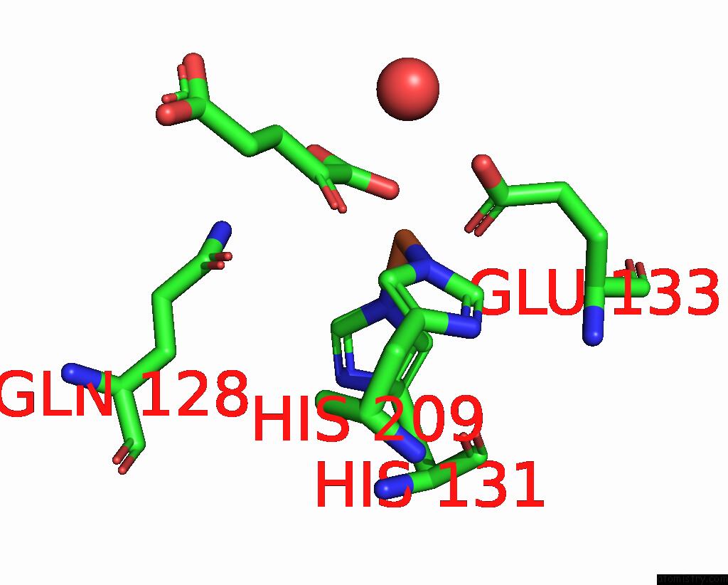
Mono view
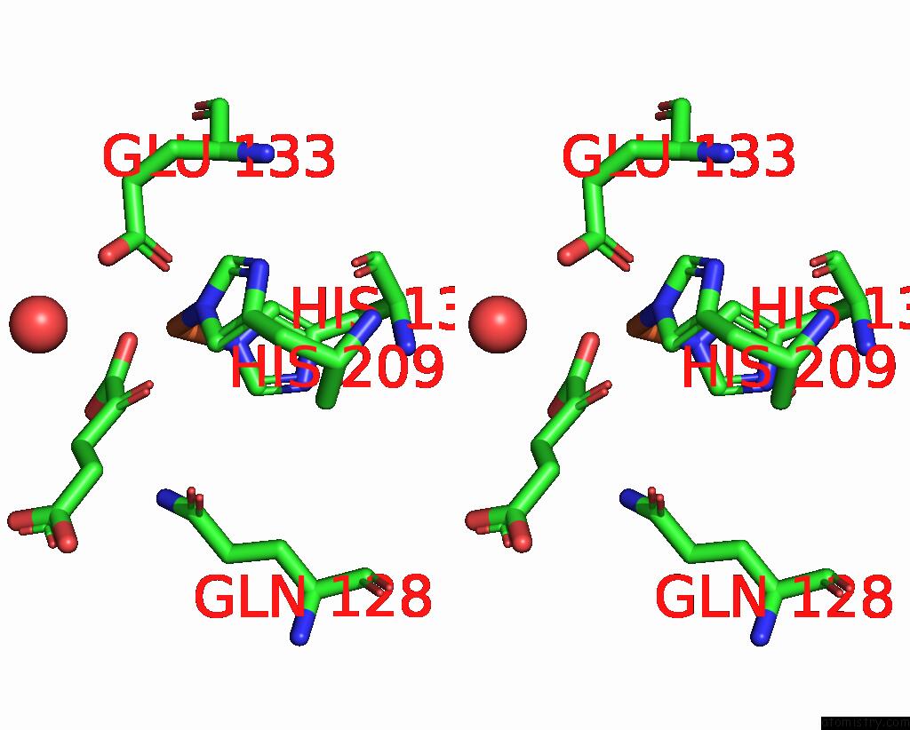
Stereo pair view
Mono view
Stereo pair view
A full contact list of Iron with other atoms in the Fe binding
site number 1 of Alpha-Ketoglutarate-Dependent Non-Heme Iron Oxygenase Eash within 5.0Å range:
|
Iron binding site 2 out of 2 in 5m0t
Go back to
Iron binding site 2 out
of 2 in the Alpha-Ketoglutarate-Dependent Non-Heme Iron Oxygenase Eash
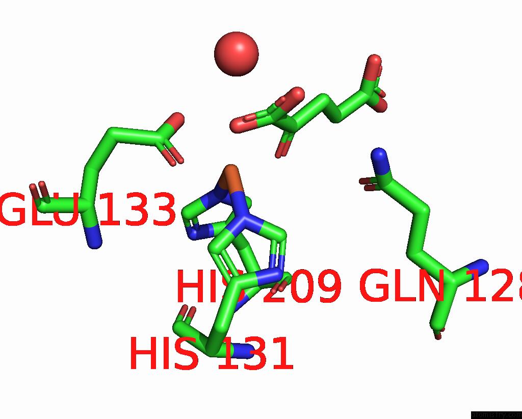
Mono view
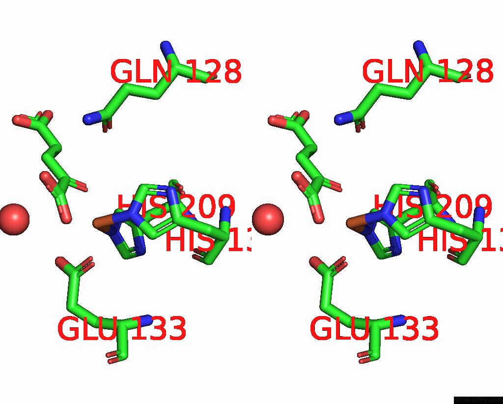
Stereo pair view
Mono view
Stereo pair view
A full contact list of Iron with other atoms in the Fe binding
site number 2 of Alpha-Ketoglutarate-Dependent Non-Heme Iron Oxygenase Eash within 5.0Å range:
|
Reference:
D.Jakubczyk,
L.Caputi,
C.E.Stevenson,
D.M.Lawson,
S.E.O'connor.
Structural Characterization of Eash (Aspergillus Japonicus) - An Oxidase Involved in Cycloclavine Biosynthesis. Chem. Commun. (Camb.) V. 52 14306 2016.
ISSN: ESSN 1364-548X
PubMed: 27885368
DOI: 10.1039/C6CC08438A
Page generated: Tue Aug 5 23:28:04 2025
ISSN: ESSN 1364-548X
PubMed: 27885368
DOI: 10.1039/C6CC08438A
Last articles
Fe in 6V93Fe in 6V59
Fe in 6UYK
Fe in 6V3R
Fe in 6UW2
Fe in 6UX0
Fe in 6UQN
Fe in 6UQM
Fe in 6UUQ
Fe in 6US6