Iron »
PDB 5m3l-5mkq »
5m3s »
Iron in PDB 5m3s: Low-Dose Fixed Target Serial Synchrotron Crystallography Structure of Metmyoglobin
Protein crystallography data
The structure of Low-Dose Fixed Target Serial Synchrotron Crystallography Structure of Metmyoglobin, PDB code: 5m3s
was solved by
D.Axford,
R.L.Owen,
D.Sherrell,
H.Muller-Werkmeister,
with X-Ray Crystallography technique. A brief refinement statistics is given in the table below:
| Resolution Low / High (Å) | 39.75 / 1.80 |
| Space group | P 6 |
| Cell size a, b, c (Å), α, β, γ (°) | 91.436, 91.436, 45.957, 90.00, 90.00, 120.00 |
| R / Rfree (%) | 18 / 22.5 |
Iron Binding Sites:
The binding sites of Iron atom in the Low-Dose Fixed Target Serial Synchrotron Crystallography Structure of Metmyoglobin
(pdb code 5m3s). This binding sites where shown within
5.0 Angstroms radius around Iron atom.
In total only one binding site of Iron was determined in the Low-Dose Fixed Target Serial Synchrotron Crystallography Structure of Metmyoglobin, PDB code: 5m3s:
In total only one binding site of Iron was determined in the Low-Dose Fixed Target Serial Synchrotron Crystallography Structure of Metmyoglobin, PDB code: 5m3s:
Iron binding site 1 out of 1 in 5m3s
Go back to
Iron binding site 1 out
of 1 in the Low-Dose Fixed Target Serial Synchrotron Crystallography Structure of Metmyoglobin
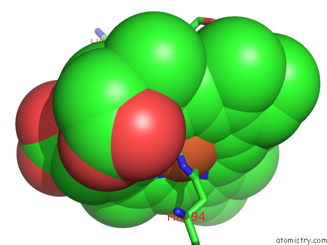
Mono view
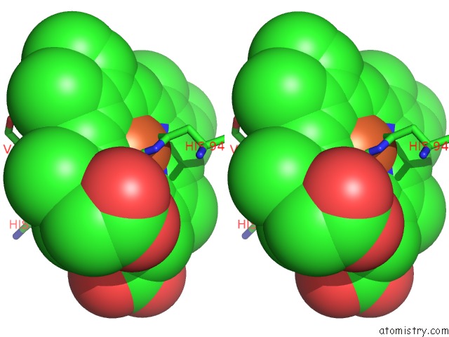
Stereo pair view
Mono view
Stereo pair view
A full contact list of Iron with other atoms in the Fe binding
site number 1 of Low-Dose Fixed Target Serial Synchrotron Crystallography Structure of Metmyoglobin within 5.0Å range:
|
Reference:
R.L.Owen,
D.Axford,
D.A.Sherrell,
A.Kuo,
O.P.Ernst,
E.C.Schulz,
R.J.Miller,
H.M.Mueller-Werkmeister.
Low-Dose Fixed-Target Serial Synchrotron Crystallography. Acta Crystallogr D Struct V. 73 373 2017BIOL.
ISSN: ISSN 2059-7983
PubMed: 28375148
DOI: 10.1107/S2059798317002996
Page generated: Tue Aug 6 05:17:13 2024
ISSN: ISSN 2059-7983
PubMed: 28375148
DOI: 10.1107/S2059798317002996
Last articles
Zn in 9J0NZn in 9J0O
Zn in 9J0P
Zn in 9FJX
Zn in 9EKB
Zn in 9C0F
Zn in 9CAH
Zn in 9CH0
Zn in 9CH3
Zn in 9CH1