Iron »
PDB 5m3l-5mkq »
5mjh »
Iron in PDB 5mjh: X-Ray Generated Oxyferrous/Water Mixed Complex of Dtpa From Streptomyces Lividans
Protein crystallography data
The structure of X-Ray Generated Oxyferrous/Water Mixed Complex of Dtpa From Streptomyces Lividans, PDB code: 5mjh
was solved by
T.Moreno Chicano,
A.K.Chaplin,
J.A.R.Worrall,
R.W.Strange,
M.A.Hough,
with X-Ray Crystallography technique. A brief refinement statistics is given in the table below:
| Resolution Low / High (Å) | 77.53 / 1.45 |
| Space group | P 1 21 1 |
| Cell size a, b, c (Å), α, β, γ (°) | 59.782, 70.627, 77.642, 90.00, 93.01, 90.00 |
| R / Rfree (%) | 22.1 / 25.4 |
Iron Binding Sites:
The binding sites of Iron atom in the X-Ray Generated Oxyferrous/Water Mixed Complex of Dtpa From Streptomyces Lividans
(pdb code 5mjh). This binding sites where shown within
5.0 Angstroms radius around Iron atom.
In total 2 binding sites of Iron where determined in the X-Ray Generated Oxyferrous/Water Mixed Complex of Dtpa From Streptomyces Lividans, PDB code: 5mjh:
Jump to Iron binding site number: 1; 2;
In total 2 binding sites of Iron where determined in the X-Ray Generated Oxyferrous/Water Mixed Complex of Dtpa From Streptomyces Lividans, PDB code: 5mjh:
Jump to Iron binding site number: 1; 2;
Iron binding site 1 out of 2 in 5mjh
Go back to
Iron binding site 1 out
of 2 in the X-Ray Generated Oxyferrous/Water Mixed Complex of Dtpa From Streptomyces Lividans
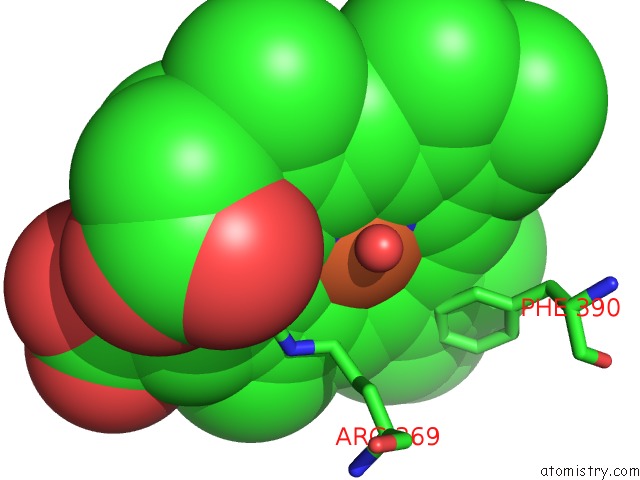
Mono view
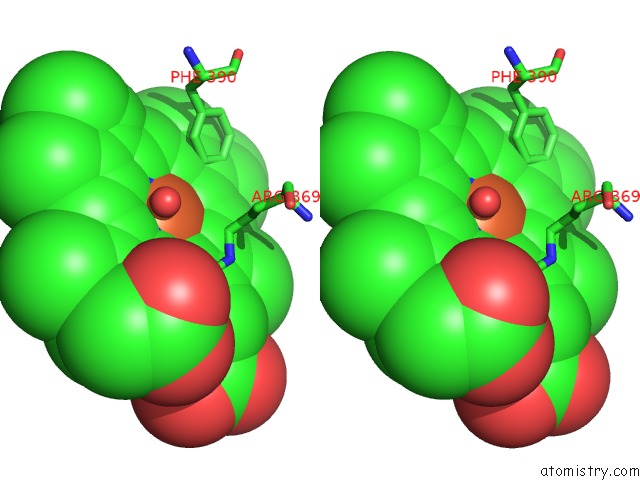
Stereo pair view
Mono view
Stereo pair view
A full contact list of Iron with other atoms in the Fe binding
site number 1 of X-Ray Generated Oxyferrous/Water Mixed Complex of Dtpa From Streptomyces Lividans within 5.0Å range:
|
Iron binding site 2 out of 2 in 5mjh
Go back to
Iron binding site 2 out
of 2 in the X-Ray Generated Oxyferrous/Water Mixed Complex of Dtpa From Streptomyces Lividans
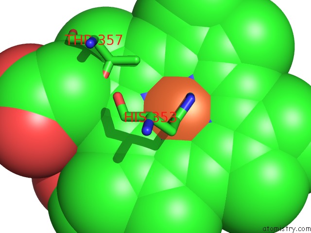
Mono view
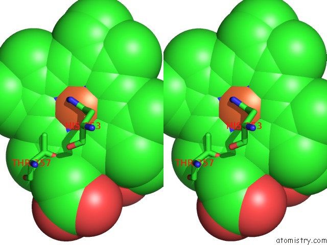
Stereo pair view
Mono view
Stereo pair view
A full contact list of Iron with other atoms in the Fe binding
site number 2 of X-Ray Generated Oxyferrous/Water Mixed Complex of Dtpa From Streptomyces Lividans within 5.0Å range:
|
Reference:
D.Kekilli,
T.Moreno-Chicano,
A.K.Chaplin,
S.Horrell,
F.S.N.Dworkowski,
J.A.R.Worrall,
R.W.Strange,
M.A.Hough.
Photoreduction and Validation of Haem-Ligand Intermediate States in Protein Crystals By in Situ Single-Crystal Spectroscopy and Diffraction. Iucrj V. 4 263 2017.
ISSN: ESSN 2052-2525
PubMed: 28512573
DOI: 10.1107/S2052252517002159
Page generated: Tue Aug 6 05:53:59 2024
ISSN: ESSN 2052-2525
PubMed: 28512573
DOI: 10.1107/S2052252517002159
Last articles
Zn in 9J0NZn in 9J0O
Zn in 9J0P
Zn in 9FJX
Zn in 9EKB
Zn in 9C0F
Zn in 9CAH
Zn in 9CH0
Zn in 9CH3
Zn in 9CH1