Iron »
PDB 5o17-5ok4 »
5o18 »
Iron in PDB 5o18: Crystal Structure of Murine Neuroglobin Mutant V140W
Protein crystallography data
The structure of Crystal Structure of Murine Neuroglobin Mutant V140W, PDB code: 5o18
was solved by
N.Colloc'h,
T.Prange,
with X-Ray Crystallography technique. A brief refinement statistics is given in the table below:
| Resolution Low / High (Å) | 20.00 / 1.86 |
| Space group | H 3 2 |
| Cell size a, b, c (Å), α, β, γ (°) | 88.563, 88.563, 116.210, 90.00, 90.00, 120.00 |
| R / Rfree (%) | 17.9 / 22.2 |
Iron Binding Sites:
The binding sites of Iron atom in the Crystal Structure of Murine Neuroglobin Mutant V140W
(pdb code 5o18). This binding sites where shown within
5.0 Angstroms radius around Iron atom.
In total only one binding site of Iron was determined in the Crystal Structure of Murine Neuroglobin Mutant V140W, PDB code: 5o18:
In total only one binding site of Iron was determined in the Crystal Structure of Murine Neuroglobin Mutant V140W, PDB code: 5o18:
Iron binding site 1 out of 1 in 5o18
Go back to
Iron binding site 1 out
of 1 in the Crystal Structure of Murine Neuroglobin Mutant V140W
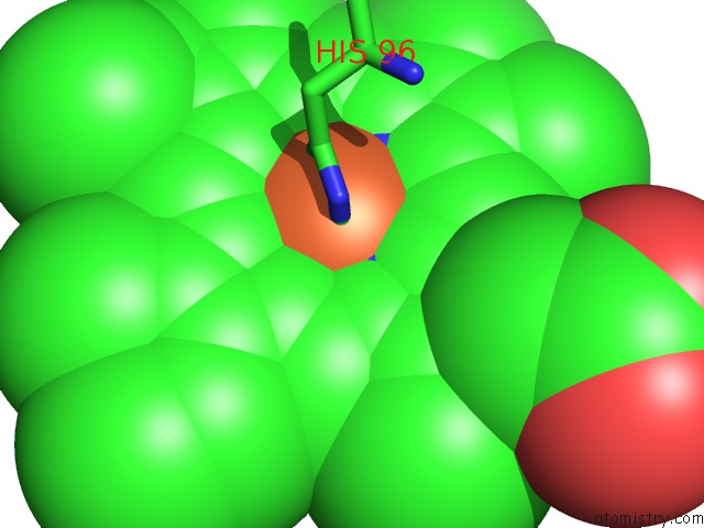
Mono view
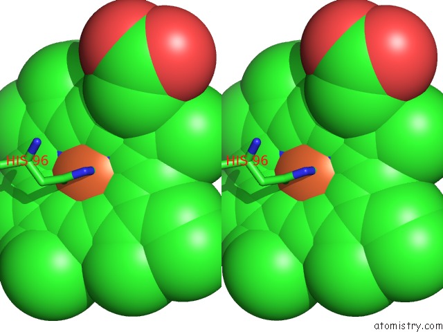
Stereo pair view
Mono view
Stereo pair view
A full contact list of Iron with other atoms in the Fe binding
site number 1 of Crystal Structure of Murine Neuroglobin Mutant V140W within 5.0Å range:
|
Reference:
N.Colloc'h,
P.Carpentier,
L.C.Montemiglio,
B.Vallone,
T.Prange.
Mapping Hydrophobic Tunnels and Cavities in Neuroglobin with Noble Gas Under Pressure. Biophys. J. V. 113 2199 2017.
ISSN: ESSN 1542-0086
PubMed: 29108649
DOI: 10.1016/J.BPJ.2017.10.014
Page generated: Tue Aug 6 06:32:51 2024
ISSN: ESSN 1542-0086
PubMed: 29108649
DOI: 10.1016/J.BPJ.2017.10.014
Last articles
Zn in 9MJ5Zn in 9HNW
Zn in 9G0L
Zn in 9FNE
Zn in 9DZN
Zn in 9E0I
Zn in 9D32
Zn in 9DAK
Zn in 8ZXC
Zn in 8ZUF