Iron »
PDB 5o17-5ok4 »
5oah »
Iron in PDB 5oah: The Periplasmic Binding Protein Ceue of Campylobacter Jejuni Binds the Iron(III) Complex of Azotochelin
Protein crystallography data
The structure of The Periplasmic Binding Protein Ceue of Campylobacter Jejuni Binds the Iron(III) Complex of Azotochelin, PDB code: 5oah
was solved by
A.D.J.Raines,
E.Blagova,
E.J.Dodson,
K.S.Wilson,
A.K.Duhme-Klair,
with X-Ray Crystallography technique. A brief refinement statistics is given in the table below:
| Resolution Low / High (Å) | 65.52 / 1.80 |
| Space group | P 1 |
| Cell size a, b, c (Å), α, β, γ (°) | 58.139, 63.410, 67.674, 82.71, 76.36, 79.03 |
| R / Rfree (%) | 20.5 / 25.1 |
Iron Binding Sites:
The binding sites of Iron atom in the The Periplasmic Binding Protein Ceue of Campylobacter Jejuni Binds the Iron(III) Complex of Azotochelin
(pdb code 5oah). This binding sites where shown within
5.0 Angstroms radius around Iron atom.
In total 2 binding sites of Iron where determined in the The Periplasmic Binding Protein Ceue of Campylobacter Jejuni Binds the Iron(III) Complex of Azotochelin, PDB code: 5oah:
Jump to Iron binding site number: 1; 2;
In total 2 binding sites of Iron where determined in the The Periplasmic Binding Protein Ceue of Campylobacter Jejuni Binds the Iron(III) Complex of Azotochelin, PDB code: 5oah:
Jump to Iron binding site number: 1; 2;
Iron binding site 1 out of 2 in 5oah
Go back to
Iron binding site 1 out
of 2 in the The Periplasmic Binding Protein Ceue of Campylobacter Jejuni Binds the Iron(III) Complex of Azotochelin
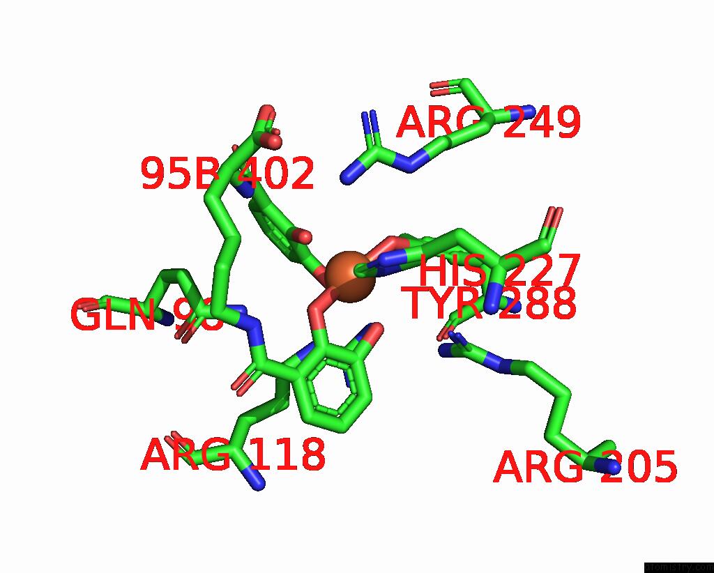
Mono view
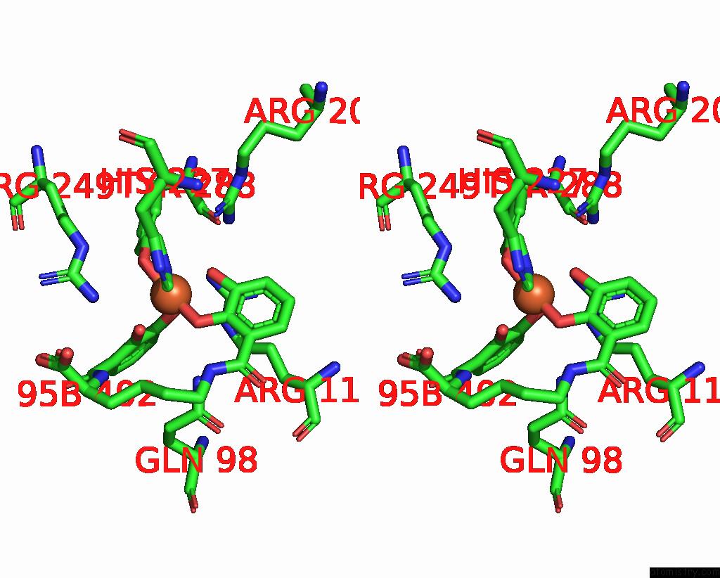
Stereo pair view
Mono view
Stereo pair view
A full contact list of Iron with other atoms in the Fe binding
site number 1 of The Periplasmic Binding Protein Ceue of Campylobacter Jejuni Binds the Iron(III) Complex of Azotochelin within 5.0Å range:
|
Iron binding site 2 out of 2 in 5oah
Go back to
Iron binding site 2 out
of 2 in the The Periplasmic Binding Protein Ceue of Campylobacter Jejuni Binds the Iron(III) Complex of Azotochelin
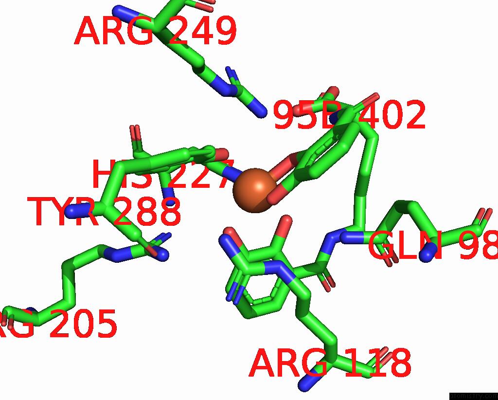
Mono view
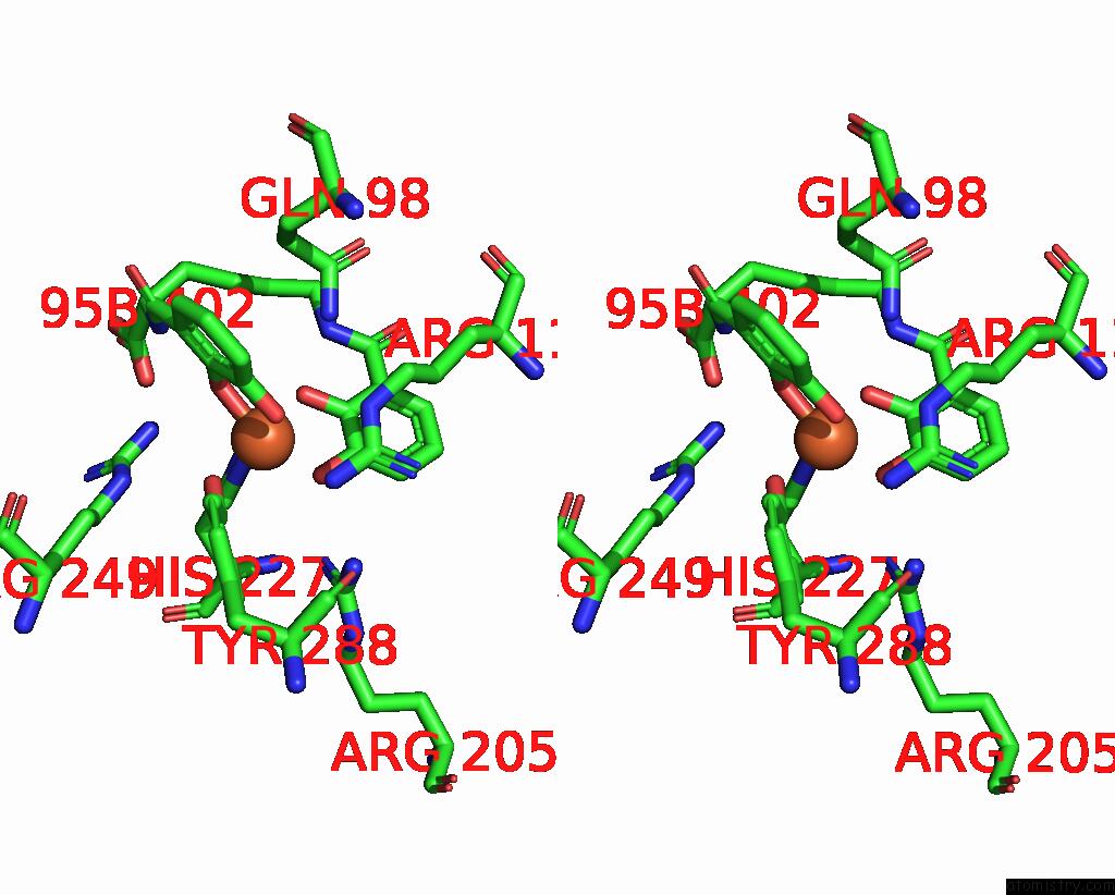
Stereo pair view
Mono view
Stereo pair view
A full contact list of Iron with other atoms in the Fe binding
site number 2 of The Periplasmic Binding Protein Ceue of Campylobacter Jejuni Binds the Iron(III) Complex of Azotochelin within 5.0Å range:
|
Reference:
A.D.J.Raines,
J.E.Clarke,
E.Blagova,
E.J.Dodson,
K.S.Wilson,
A.K.Duhme-Klair.
Redox-Switchable Siderophore Anchor Enables Reversible Artificial Metalloenzyme Assembly Nat Catal 2018.
DOI: 10.1038/S41929-018-0124-3
Page generated: Tue Aug 6 06:34:33 2024
DOI: 10.1038/S41929-018-0124-3
Last articles
F in 4JSXF in 4JSV
F in 4JTQ
F in 4JSC
F in 4JSJ
F in 4JSM
F in 4JQG
F in 4JSI
F in 4JQ2
F in 4JP4