Iron in PDB 5of4: The Cryo-Em Structure of Human Tfiih
Enzymatic activity of The Cryo-Em Structure of Human Tfiih
All present enzymatic activity of The Cryo-Em Structure of Human Tfiih:
3.6.4.12;
Iron Binding Sites:
The binding sites of Iron atom in the The Cryo-Em Structure of Human Tfiih
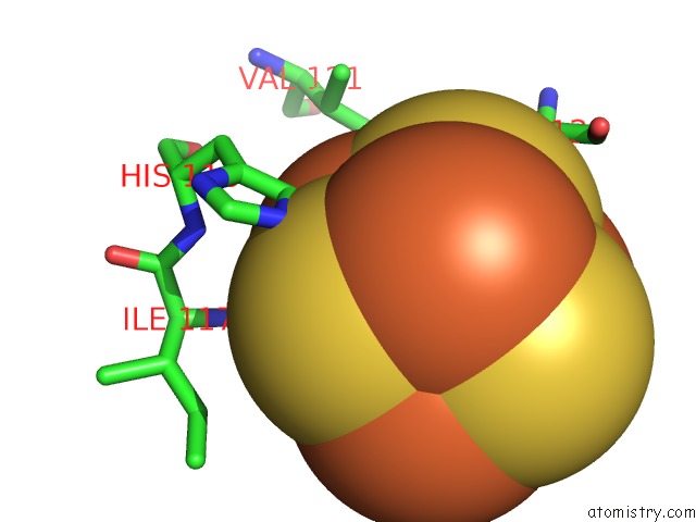
(pdb code 5of4). This binding sites where shown within
5.0 Angstroms radius around Iron atom.
In total 4 binding sites of Iron where determined in the
The Cryo-Em Structure of Human Tfiih, PDB code: 5of4:
Jump to Iron binding site number:
1;
2;
3;
4;
Iron binding site 1 out
of 4 in 5of4
Go back to
Iron Binding Sites List in 5of4
Iron binding site 1 out
of 4 in the The Cryo-Em Structure of Human Tfiih
Mono view
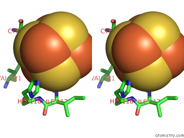
Stereo pair view
|
A full contact list of Iron with other atoms in the Fe binding
site number 1 of The Cryo-Em Structure of Human Tfiih within 5.0Å range:
probe
|
atom
|
residue
|
distance (Å)
|
B
|
Occ
|
B:Fe1000
b:97.0
occ:1.00
|
FE1
|
B:SF41000
|
0.0
|
97.0
|
1.0
|
S4
|
B:SF41000
|
2.1
|
0.6
|
1.0
|
S2
|
B:SF41000
|
2.1
|
95.2
|
1.0
|
S3
|
B:SF41000
|
2.2
|
0.7
|
1.0
|
SG
|
B:CYS116
|
2.2
|
0.2
|
1.0
|
FE3
|
B:SF41000
|
2.9
|
0.4
|
1.0
|
FE2
|
B:SF41000
|
2.9
|
0.3
|
1.0
|
CB
|
B:CYS116
|
3.0
|
0.5
|
1.0
|
FE4
|
B:SF41000
|
3.3
|
0.8
|
1.0
|
CA
|
B:CYS116
|
3.6
|
0.6
|
1.0
|
CD2
|
B:HIS118
|
3.7
|
0.2
|
1.0
|
S1
|
B:SF41000
|
3.8
|
0.6
|
1.0
|
SG
|
B:CYS134
|
4.2
|
0.2
|
1.0
|
NE2
|
B:HIS118
|
4.3
|
0.9
|
1.0
|
N
|
B:ILE117
|
4.5
|
1.0
|
1.0
|
C
|
B:CYS116
|
4.6
|
0.5
|
1.0
|
N
|
B:CYS116
|
4.6
|
0.9
|
1.0
|
CG
|
B:HIS118
|
4.7
|
0.3
|
1.0
|
O
|
B:LEU115
|
4.7
|
0.4
|
1.0
|
CB
|
B:CYS134
|
4.8
|
0.2
|
1.0
|
C
|
B:LEU115
|
5.0
|
0.8
|
1.0
|
CG1
|
B:VAL121
|
5.0
|
0.2
|
1.0
|
|
Iron binding site 2 out
of 4 in 5of4
Go back to
Iron Binding Sites List in 5of4
Iron binding site 2 out
of 4 in the The Cryo-Em Structure of Human Tfiih
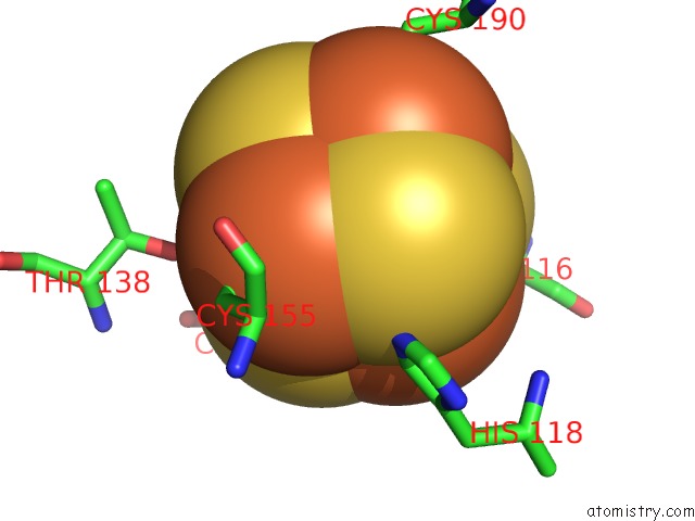
Mono view
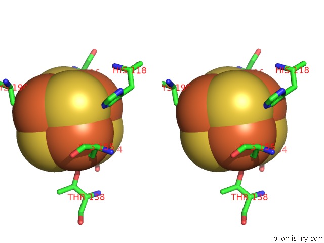
Stereo pair view
|
A full contact list of Iron with other atoms in the Fe binding
site number 2 of The Cryo-Em Structure of Human Tfiih within 5.0Å range:
probe
|
atom
|
residue
|
distance (Å)
|
B
|
Occ
|
B:Fe1000
b:0.3
occ:1.00
|
FE2
|
B:SF41000
|
0.0
|
0.3
|
1.0
|
S3
|
B:SF41000
|
2.1
|
0.7
|
1.0
|
S4
|
B:SF41000
|
2.2
|
0.6
|
1.0
|
S1
|
B:SF41000
|
2.2
|
0.6
|
1.0
|
SG
|
B:CYS155
|
2.3
|
0.7
|
1.0
|
FE1
|
B:SF41000
|
2.9
|
97.0
|
1.0
|
FE4
|
B:SF41000
|
3.0
|
0.8
|
1.0
|
FE3
|
B:SF41000
|
3.0
|
0.4
|
1.0
|
S2
|
B:SF41000
|
3.6
|
95.2
|
1.0
|
CB
|
B:CYS155
|
3.8
|
0.1
|
1.0
|
OG1
|
B:THR138
|
3.9
|
0.2
|
1.0
|
NE2
|
B:HIS118
|
4.2
|
0.9
|
1.0
|
SG
|
B:CYS134
|
4.2
|
0.2
|
1.0
|
CD2
|
B:HIS118
|
4.3
|
0.2
|
1.0
|
CA
|
B:CYS155
|
4.5
|
0.6
|
1.0
|
SG
|
B:CYS190
|
4.7
|
0.1
|
1.0
|
SG
|
B:CYS116
|
4.9
|
0.2
|
1.0
|
|
Iron binding site 3 out
of 4 in 5of4
Go back to
Iron Binding Sites List in 5of4
Iron binding site 3 out
of 4 in the The Cryo-Em Structure of Human Tfiih
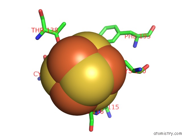
Mono view
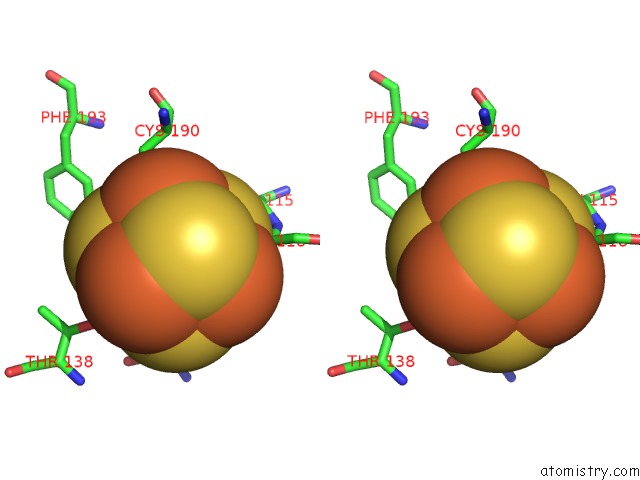
Stereo pair view
|
A full contact list of Iron with other atoms in the Fe binding
site number 3 of The Cryo-Em Structure of Human Tfiih within 5.0Å range:
probe
|
atom
|
residue
|
distance (Å)
|
B
|
Occ
|
B:Fe1000
b:0.4
occ:1.00
|
FE3
|
B:SF41000
|
0.0
|
0.4
|
1.0
|
S1
|
B:SF41000
|
2.1
|
0.6
|
1.0
|
S4
|
B:SF41000
|
2.2
|
0.6
|
1.0
|
S2
|
B:SF41000
|
2.3
|
95.2
|
1.0
|
SG
|
B:CYS134
|
2.3
|
0.2
|
1.0
|
FE1
|
B:SF41000
|
2.9
|
97.0
|
1.0
|
FE4
|
B:SF41000
|
2.9
|
0.8
|
1.0
|
FE2
|
B:SF41000
|
3.0
|
0.3
|
1.0
|
S3
|
B:SF41000
|
3.6
|
0.7
|
1.0
|
CB
|
B:CYS134
|
3.8
|
0.2
|
1.0
|
CD1
|
B:LEU115
|
4.2
|
0.6
|
1.0
|
CG
|
B:LEU115
|
4.3
|
0.8
|
1.0
|
CD2
|
B:LEU115
|
4.7
|
93.3
|
1.0
|
CE2
|
B:PHE193
|
4.8
|
0.5
|
1.0
|
OG1
|
B:THR138
|
4.8
|
0.2
|
1.0
|
SG
|
B:CYS116
|
4.9
|
0.2
|
1.0
|
CA
|
B:CYS134
|
4.9
|
0.5
|
1.0
|
SG
|
B:CYS190
|
5.0
|
0.1
|
1.0
|
CB
|
B:CYS116
|
5.0
|
0.5
|
1.0
|
|
Iron binding site 4 out
of 4 in 5of4
Go back to
Iron Binding Sites List in 5of4
Iron binding site 4 out
of 4 in the The Cryo-Em Structure of Human Tfiih
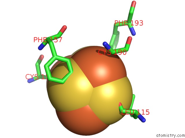
Mono view
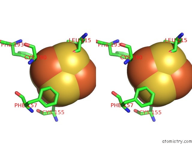
Stereo pair view
|
A full contact list of Iron with other atoms in the Fe binding
site number 4 of The Cryo-Em Structure of Human Tfiih within 5.0Å range:
probe
|
atom
|
residue
|
distance (Å)
|
B
|
Occ
|
B:Fe1000
b:0.8
occ:1.00
|
FE4
|
B:SF41000
|
0.0
|
0.8
|
1.0
|
S2
|
B:SF41000
|
2.1
|
95.2
|
1.0
|
S1
|
B:SF41000
|
2.1
|
0.6
|
1.0
|
SG
|
B:CYS190
|
2.2
|
0.1
|
1.0
|
S3
|
B:SF41000
|
2.2
|
0.7
|
1.0
|
FE3
|
B:SF41000
|
2.9
|
0.4
|
1.0
|
FE2
|
B:SF41000
|
3.0
|
0.3
|
1.0
|
FE1
|
B:SF41000
|
3.3
|
97.0
|
1.0
|
CB
|
B:CYS190
|
3.6
|
96.4
|
1.0
|
S4
|
B:SF41000
|
3.8
|
0.6
|
1.0
|
CE2
|
B:PHE157
|
4.1
|
0.4
|
1.0
|
CA
|
B:CYS190
|
4.4
|
0.1
|
1.0
|
O
|
B:LEU115
|
4.5
|
0.4
|
1.0
|
CE2
|
B:PHE193
|
4.5
|
0.5
|
1.0
|
CD2
|
B:PHE157
|
4.6
|
0.5
|
1.0
|
CD2
|
B:PHE193
|
4.7
|
0.5
|
1.0
|
SG
|
B:CYS155
|
4.8
|
0.7
|
1.0
|
CZ
|
B:PHE157
|
4.9
|
0.9
|
1.0
|
CD1
|
B:LEU115
|
4.9
|
0.6
|
1.0
|
|
Reference:
B.J.Greber,
T.H.D.Nguyen,
J.Fang,
P.V.Afonine,
P.D.Adams,
E.Nogales.
The Cryo-Electron Microscopy Structure of Human Transcription Factor Iih. Nature V. 549 414 2017.
ISSN: ESSN 1476-4687
PubMed: 28902838
DOI: 10.1038/NATURE23903
Page generated: Tue Aug 6 06:57:06 2024
|