Iron »
PDB 5okd-5sx2 »
5omr »
Iron in PDB 5omr: Crystal Structure of Amycolatopsis Cytochrome P450 Gcoa in Complex with Vanillin.
Protein crystallography data
The structure of Crystal Structure of Amycolatopsis Cytochrome P450 Gcoa in Complex with Vanillin., PDB code: 5omr
was solved by
S.J.B.Mallinson,
C.W.Johnson,
E.L.Neidle,
G.T.Beckham,
J.E.Mcgeehan,
with X-Ray Crystallography technique. A brief refinement statistics is given in the table below:
| Resolution Low / High (Å) | 36.81 / 1.68 |
| Space group | P 43 21 2 |
| Cell size a, b, c (Å), α, β, γ (°) | 104.110, 104.110, 115.660, 90.00, 90.00, 90.00 |
| R / Rfree (%) | 14.7 / 17 |
Iron Binding Sites:
The binding sites of Iron atom in the Crystal Structure of Amycolatopsis Cytochrome P450 Gcoa in Complex with Vanillin.
(pdb code 5omr). This binding sites where shown within
5.0 Angstroms radius around Iron atom.
In total only one binding site of Iron was determined in the Crystal Structure of Amycolatopsis Cytochrome P450 Gcoa in Complex with Vanillin., PDB code: 5omr:
In total only one binding site of Iron was determined in the Crystal Structure of Amycolatopsis Cytochrome P450 Gcoa in Complex with Vanillin., PDB code: 5omr:
Iron binding site 1 out of 1 in 5omr
Go back to
Iron binding site 1 out
of 1 in the Crystal Structure of Amycolatopsis Cytochrome P450 Gcoa in Complex with Vanillin.

Mono view
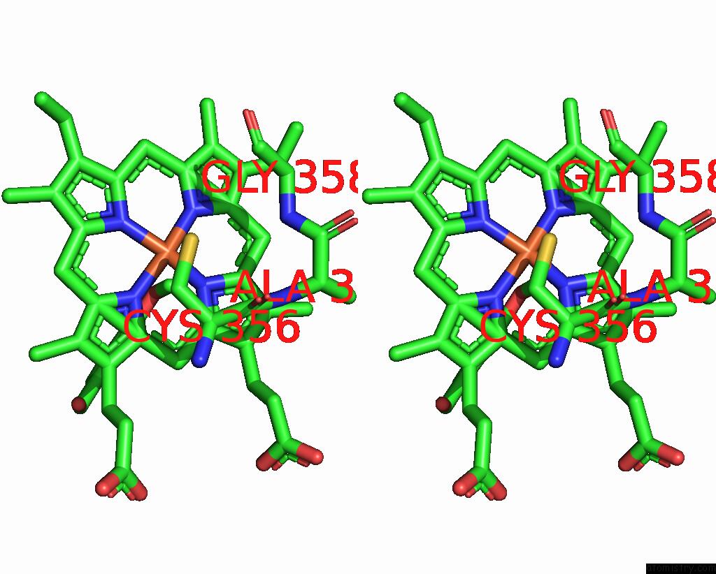
Stereo pair view
Mono view
Stereo pair view
A full contact list of Iron with other atoms in the Fe binding
site number 1 of Crystal Structure of Amycolatopsis Cytochrome P450 Gcoa in Complex with Vanillin. within 5.0Å range:
|
Reference:
S.J.B.Mallinson,
M.M.Machovina,
R.L.Silveira,
M.Garcia-Borras,
N.Gallup,
C.W.Johnson,
M.D.Allen,
M.S.Skaf,
M.F.Crowley,
E.L.Neidle,
K.N.Houk,
G.T.Beckham,
J.L.Dubois,
J.E.Mcgeehan.
A Promiscuous Cytochrome P450 Aromatic O-Demethylase For Lignin Bioconversion. Nat Commun V. 9 2487 2018.
ISSN: ESSN 2041-1723
PubMed: 29950589
DOI: 10.1038/S41467-018-04878-2
Page generated: Tue Aug 6 08:26:45 2024
ISSN: ESSN 2041-1723
PubMed: 29950589
DOI: 10.1038/S41467-018-04878-2
Last articles
Zn in 9J0NZn in 9J0O
Zn in 9J0P
Zn in 9FJX
Zn in 9EKB
Zn in 9C0F
Zn in 9CAH
Zn in 9CH0
Zn in 9CH3
Zn in 9CH1