Iron »
PDB 5sx3-5tia »
5sxw »
Iron in PDB 5sxw: Crystal Structure of the E198A Variant of Catalase-Peroxidase Katg of Burkholderia Pseudomallei
Enzymatic activity of Crystal Structure of the E198A Variant of Catalase-Peroxidase Katg of Burkholderia Pseudomallei
All present enzymatic activity of Crystal Structure of the E198A Variant of Catalase-Peroxidase Katg of Burkholderia Pseudomallei:
1.11.1.21;
1.11.1.21;
Protein crystallography data
The structure of Crystal Structure of the E198A Variant of Catalase-Peroxidase Katg of Burkholderia Pseudomallei, PDB code: 5sxw
was solved by
P.C.Loewen,
with X-Ray Crystallography technique. A brief refinement statistics is given in the table below:
| Resolution Low / High (Å) | 20.00 / 1.60 |
| Space group | P 21 21 21 |
| Cell size a, b, c (Å), α, β, γ (°) | 100.340, 116.020, 174.710, 90.00, 90.00, 90.00 |
| R / Rfree (%) | 14.3 / 16.9 |
Other elements in 5sxw:
The structure of Crystal Structure of the E198A Variant of Catalase-Peroxidase Katg of Burkholderia Pseudomallei also contains other interesting chemical elements:
| Sodium | (Na) | 2 atoms |
Iron Binding Sites:
The binding sites of Iron atom in the Crystal Structure of the E198A Variant of Catalase-Peroxidase Katg of Burkholderia Pseudomallei
(pdb code 5sxw). This binding sites where shown within
5.0 Angstroms radius around Iron atom.
In total 2 binding sites of Iron where determined in the Crystal Structure of the E198A Variant of Catalase-Peroxidase Katg of Burkholderia Pseudomallei, PDB code: 5sxw:
Jump to Iron binding site number: 1; 2;
In total 2 binding sites of Iron where determined in the Crystal Structure of the E198A Variant of Catalase-Peroxidase Katg of Burkholderia Pseudomallei, PDB code: 5sxw:
Jump to Iron binding site number: 1; 2;
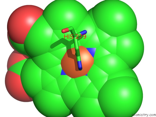
Iron binding site 1 out of 2 in 5sxw
Go back to
Iron binding site 1 out
of 2 in the Crystal Structure of the E198A Variant of Catalase-Peroxidase Katg of Burkholderia Pseudomallei

Mono view

Stereo pair view
Mono view
Stereo pair view
A full contact list of Iron with other atoms in the Fe binding
site number 1 of Crystal Structure of the E198A Variant of Catalase-Peroxidase Katg of Burkholderia Pseudomallei within 5.0Å range:
|
Iron binding site 2 out of 2 in 5sxw
Go back to
Iron binding site 2 out
of 2 in the Crystal Structure of the E198A Variant of Catalase-Peroxidase Katg of Burkholderia Pseudomallei
Mono view

Stereo pair view
Mono view
Stereo pair view
A full contact list of Iron with other atoms in the Fe binding
site number 2 of Crystal Structure of the E198A Variant of Catalase-Peroxidase Katg of Burkholderia Pseudomallei within 5.0Å range:
|
Reference:
B.Wiseman,
X.Carpena,
M.Feliz,
L.J.Donald,
M.Pons,
I.Fita,
P.C.Loewen.
Isonicotinic Acid Hydrazide Conversion to Isonicotinyl-Nad By Catalase-Peroxidases. J. Biol. Chem. V. 285 26662 2010.
ISSN: ESSN 1083-351X
PubMed: 20554537
DOI: 10.1074/JBC.M110.139428
Page generated: Tue Aug 6 08:48:37 2024
ISSN: ESSN 1083-351X
PubMed: 20554537
DOI: 10.1074/JBC.M110.139428
Last articles
Fe in 2YXOFe in 2YRS
Fe in 2YXC
Fe in 2YNM
Fe in 2YVJ
Fe in 2YP1
Fe in 2YU2
Fe in 2YU1
Fe in 2YQB
Fe in 2YOO