Iron »
PDB 5sx3-5tia »
5t6q »
Iron in PDB 5t6q: Structure of Cytochrome P450 4B1 (CYP4B1) Complexed with Octane: An N- Alkane and Fatty Acid Omega-Hydroxylase with A Covalently Bound Heme
Enzymatic activity of Structure of Cytochrome P450 4B1 (CYP4B1) Complexed with Octane: An N- Alkane and Fatty Acid Omega-Hydroxylase with A Covalently Bound Heme
All present enzymatic activity of Structure of Cytochrome P450 4B1 (CYP4B1) Complexed with Octane: An N- Alkane and Fatty Acid Omega-Hydroxylase with A Covalently Bound Heme:
1.14.14.1;
1.14.14.1;
Protein crystallography data
The structure of Structure of Cytochrome P450 4B1 (CYP4B1) Complexed with Octane: An N- Alkane and Fatty Acid Omega-Hydroxylase with A Covalently Bound Heme, PDB code: 5t6q
was solved by
M.Hsu,
E.F.Johnson,
with X-Ray Crystallography technique. A brief refinement statistics is given in the table below:
| Resolution Low / High (Å) | 38.37 / 2.70 |
| Space group | P 32 2 1 |
| Cell size a, b, c (Å), α, β, γ (°) | 109.678, 109.678, 125.817, 90.00, 90.00, 120.00 |
| R / Rfree (%) | 20 / 24 |
Iron Binding Sites:
The binding sites of Iron atom in the Structure of Cytochrome P450 4B1 (CYP4B1) Complexed with Octane: An N- Alkane and Fatty Acid Omega-Hydroxylase with A Covalently Bound Heme
(pdb code 5t6q). This binding sites where shown within
5.0 Angstroms radius around Iron atom.
In total only one binding site of Iron was determined in the Structure of Cytochrome P450 4B1 (CYP4B1) Complexed with Octane: An N- Alkane and Fatty Acid Omega-Hydroxylase with A Covalently Bound Heme, PDB code: 5t6q:
In total only one binding site of Iron was determined in the Structure of Cytochrome P450 4B1 (CYP4B1) Complexed with Octane: An N- Alkane and Fatty Acid Omega-Hydroxylase with A Covalently Bound Heme, PDB code: 5t6q:
Iron binding site 1 out of 1 in 5t6q
Go back to
Iron binding site 1 out
of 1 in the Structure of Cytochrome P450 4B1 (CYP4B1) Complexed with Octane: An N- Alkane and Fatty Acid Omega-Hydroxylase with A Covalently Bound Heme
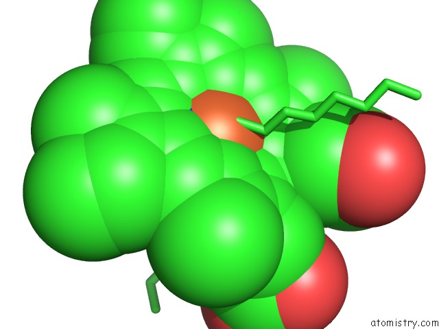
Mono view
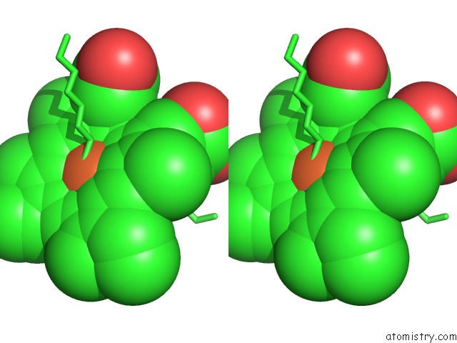
Stereo pair view
Mono view
Stereo pair view
A full contact list of Iron with other atoms in the Fe binding
site number 1 of Structure of Cytochrome P450 4B1 (CYP4B1) Complexed with Octane: An N- Alkane and Fatty Acid Omega-Hydroxylase with A Covalently Bound Heme within 5.0Å range:
|
Reference:
M.H.Hsu,
B.R.Baer,
A.E.Rettie,
E.F.Johnson.
The Crystal Structure of Cytochrome P450 4B1 (CYP4B1) Monooxygenase Complexed with Octane Discloses Several Structural Adaptations For Omega-Hydroxylation. J. Biol. Chem. V. 292 5610 2017.
ISSN: ESSN 1083-351X
PubMed: 28167536
DOI: 10.1074/JBC.M117.775494
Page generated: Tue Aug 6 08:54:27 2024
ISSN: ESSN 1083-351X
PubMed: 28167536
DOI: 10.1074/JBC.M117.775494
Last articles
Fe in 2YXOFe in 2YRS
Fe in 2YXC
Fe in 2YNM
Fe in 2YVJ
Fe in 2YP1
Fe in 2YU2
Fe in 2YU1
Fe in 2YQB
Fe in 2YOO