Iron »
PDB 5sx3-5tia »
5tgs »
Iron in PDB 5tgs: Crystal Structure of Quee From Bacillus Subtilis with Methionine Bound
Enzymatic activity of Crystal Structure of Quee From Bacillus Subtilis with Methionine Bound
All present enzymatic activity of Crystal Structure of Quee From Bacillus Subtilis with Methionine Bound:
4.3.99.3;
4.3.99.3;
Protein crystallography data
The structure of Crystal Structure of Quee From Bacillus Subtilis with Methionine Bound, PDB code: 5tgs
was solved by
T.A.J.Grell,
D.P.Dowling,
C.L.Drennan,
with X-Ray Crystallography technique. A brief refinement statistics is given in the table below:
| Resolution Low / High (Å) | 42.01 / 2.55 |
| Space group | P 21 21 21 |
| Cell size a, b, c (Å), α, β, γ (°) | 54.182, 79.297, 122.224, 90.00, 90.00, 90.00 |
| R / Rfree (%) | 21.6 / 24.8 |
Iron Binding Sites:
The binding sites of Iron atom in the Crystal Structure of Quee From Bacillus Subtilis with Methionine Bound
(pdb code 5tgs). This binding sites where shown within
5.0 Angstroms radius around Iron atom.
In total 8 binding sites of Iron where determined in the Crystal Structure of Quee From Bacillus Subtilis with Methionine Bound, PDB code: 5tgs:
Jump to Iron binding site number: 1; 2; 3; 4; 5; 6; 7; 8;
In total 8 binding sites of Iron where determined in the Crystal Structure of Quee From Bacillus Subtilis with Methionine Bound, PDB code: 5tgs:
Jump to Iron binding site number: 1; 2; 3; 4; 5; 6; 7; 8;
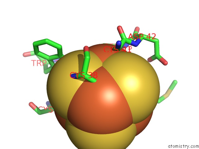
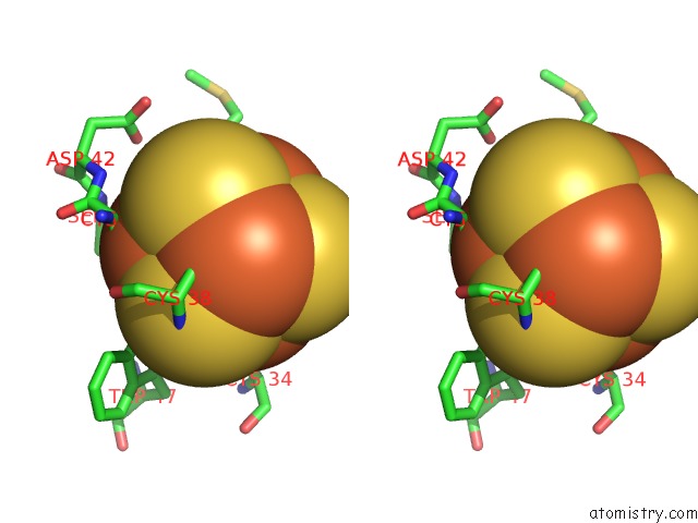
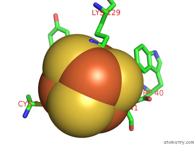
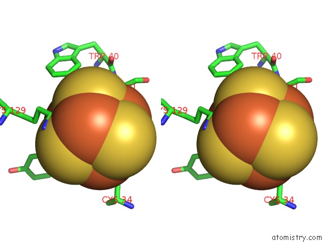
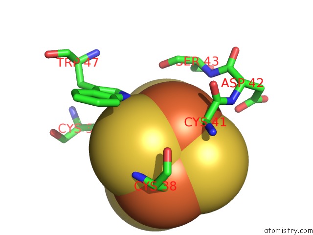
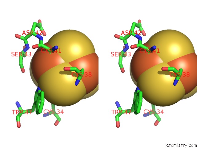
Iron binding site 1 out of 8 in 5tgs
Go back to
Iron binding site 1 out
of 8 in the Crystal Structure of Quee From Bacillus Subtilis with Methionine Bound
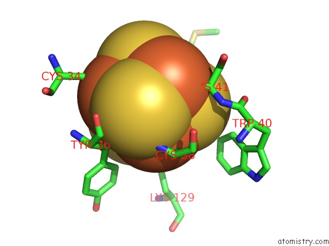
Mono view
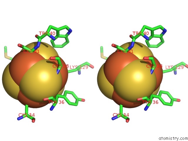
Stereo pair view
Mono view
Stereo pair view
A full contact list of Iron with other atoms in the Fe binding
site number 1 of Crystal Structure of Quee From Bacillus Subtilis with Methionine Bound within 5.0Å range:
|
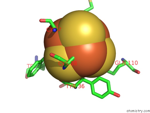
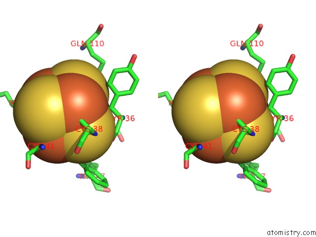
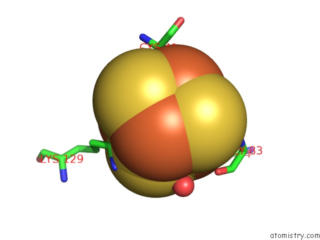
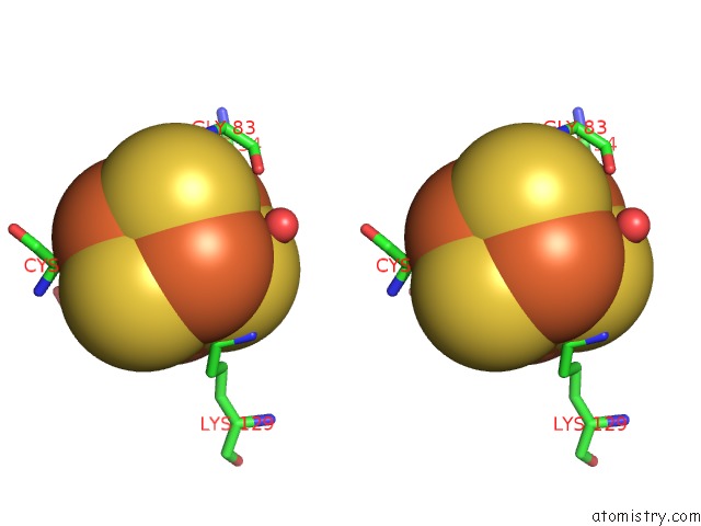
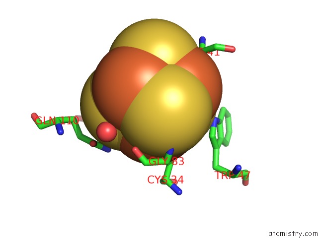
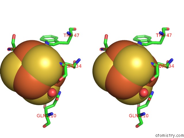
Iron binding site 2 out of 8 in 5tgs
Go back to
Iron binding site 2 out
of 8 in the Crystal Structure of Quee From Bacillus Subtilis with Methionine Bound
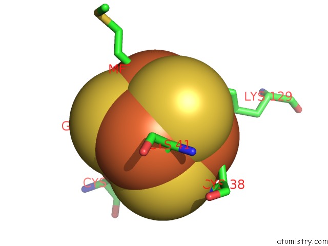
Mono view
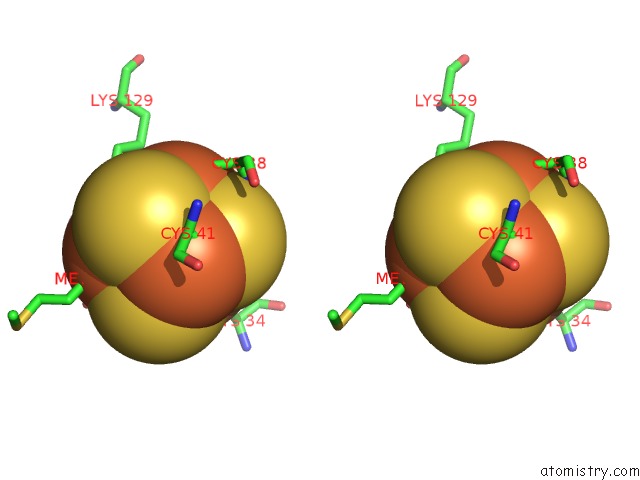
Stereo pair view
Mono view
Stereo pair view
A full contact list of Iron with other atoms in the Fe binding
site number 2 of Crystal Structure of Quee From Bacillus Subtilis with Methionine Bound within 5.0Å range:
|
Iron binding site 3 out of 8 in 5tgs
Go back to
Iron binding site 3 out
of 8 in the Crystal Structure of Quee From Bacillus Subtilis with Methionine Bound
Mono view
Stereo pair view
Mono view
Stereo pair view
A full contact list of Iron with other atoms in the Fe binding
site number 3 of Crystal Structure of Quee From Bacillus Subtilis with Methionine Bound within 5.0Å range:
|
Iron binding site 4 out of 8 in 5tgs
Go back to
Iron binding site 4 out
of 8 in the Crystal Structure of Quee From Bacillus Subtilis with Methionine Bound
Mono view
Stereo pair view
Mono view
Stereo pair view
A full contact list of Iron with other atoms in the Fe binding
site number 4 of Crystal Structure of Quee From Bacillus Subtilis with Methionine Bound within 5.0Å range:
|
Iron binding site 5 out of 8 in 5tgs
Go back to
Iron binding site 5 out
of 8 in the Crystal Structure of Quee From Bacillus Subtilis with Methionine Bound
Mono view
Stereo pair view
Mono view
Stereo pair view
A full contact list of Iron with other atoms in the Fe binding
site number 5 of Crystal Structure of Quee From Bacillus Subtilis with Methionine Bound within 5.0Å range:
|
Iron binding site 6 out of 8 in 5tgs
Go back to
Iron binding site 6 out
of 8 in the Crystal Structure of Quee From Bacillus Subtilis with Methionine Bound
Mono view
Stereo pair view
Mono view
Stereo pair view
A full contact list of Iron with other atoms in the Fe binding
site number 6 of Crystal Structure of Quee From Bacillus Subtilis with Methionine Bound within 5.0Å range:
|
Iron binding site 7 out of 8 in 5tgs
Go back to
Iron binding site 7 out
of 8 in the Crystal Structure of Quee From Bacillus Subtilis with Methionine Bound
Mono view
Stereo pair view
Mono view
Stereo pair view
A full contact list of Iron with other atoms in the Fe binding
site number 7 of Crystal Structure of Quee From Bacillus Subtilis with Methionine Bound within 5.0Å range:
|
Iron binding site 8 out of 8 in 5tgs
Go back to
Iron binding site 8 out
of 8 in the Crystal Structure of Quee From Bacillus Subtilis with Methionine Bound
Mono view
Stereo pair view
Mono view
Stereo pair view
A full contact list of Iron with other atoms in the Fe binding
site number 8 of Crystal Structure of Quee From Bacillus Subtilis with Methionine Bound within 5.0Å range:
|
Reference:
N.A.Bruender,
T.A.Grell,
D.P.Dowling,
R.M.Mccarty,
C.L.Drennan,
V.Bandarian.
7-Carboxy-7-Deazaguanine Synthase: A Radical S-Adenosyl-L-Methionine Enzyme with Polar Tendencies. J. Am. Chem. Soc. V. 139 1912 2017.
ISSN: ESSN 1520-5126
PubMed: 28045519
DOI: 10.1021/JACS.6B11381
Page generated: Tue Aug 6 08:59:43 2024
ISSN: ESSN 1520-5126
PubMed: 28045519
DOI: 10.1021/JACS.6B11381
Last articles
Zn in 9MJ5Zn in 9HNW
Zn in 9G0L
Zn in 9FNE
Zn in 9DZN
Zn in 9E0I
Zn in 9D32
Zn in 9DAK
Zn in 8ZXC
Zn in 8ZUF