Iron »
PDB 5tis-5ufj »
5u3i »
Iron in PDB 5u3i: Crystal Structure of Carbonmonoxy Hemoglobin S (Liganded Sickle Cell Hemoglobin) Complexed with Gbt Compound 31
Protein crystallography data
The structure of Crystal Structure of Carbonmonoxy Hemoglobin S (Liganded Sickle Cell Hemoglobin) Complexed with Gbt Compound 31, PDB code: 5u3i
was solved by
J.R.Partridge,
R.M.Choy,
Z.Li,
B.Metcalf,
with X-Ray Crystallography technique. A brief refinement statistics is given in the table below:
| Resolution Low / High (Å) | 29.81 / 1.95 |
| Space group | P 21 21 21 |
| Cell size a, b, c (Å), α, β, γ (°) | 57.385, 59.040, 172.637, 90.00, 90.00, 90.00 |
| R / Rfree (%) | 20.6 / 25.3 |
Iron Binding Sites:
The binding sites of Iron atom in the Crystal Structure of Carbonmonoxy Hemoglobin S (Liganded Sickle Cell Hemoglobin) Complexed with Gbt Compound 31
(pdb code 5u3i). This binding sites where shown within
5.0 Angstroms radius around Iron atom.
In total 4 binding sites of Iron where determined in the Crystal Structure of Carbonmonoxy Hemoglobin S (Liganded Sickle Cell Hemoglobin) Complexed with Gbt Compound 31, PDB code: 5u3i:
Jump to Iron binding site number: 1; 2; 3; 4;
In total 4 binding sites of Iron where determined in the Crystal Structure of Carbonmonoxy Hemoglobin S (Liganded Sickle Cell Hemoglobin) Complexed with Gbt Compound 31, PDB code: 5u3i:
Jump to Iron binding site number: 1; 2; 3; 4;
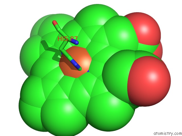
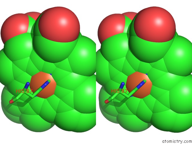
Iron binding site 1 out of 4 in 5u3i
Go back to
Iron binding site 1 out
of 4 in the Crystal Structure of Carbonmonoxy Hemoglobin S (Liganded Sickle Cell Hemoglobin) Complexed with Gbt Compound 31
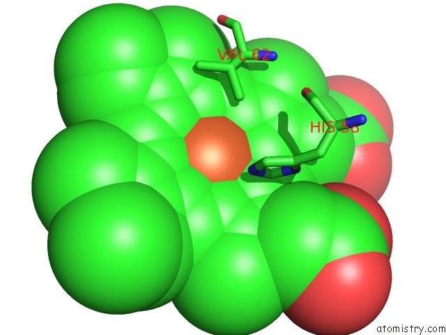
Mono view
Stereo pair view
Mono view
Stereo pair view
A full contact list of Iron with other atoms in the Fe binding
site number 1 of Crystal Structure of Carbonmonoxy Hemoglobin S (Liganded Sickle Cell Hemoglobin) Complexed with Gbt Compound 31 within 5.0Å range:
|
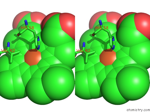
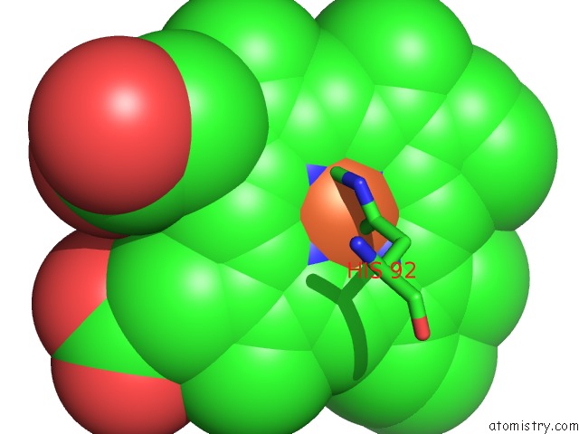
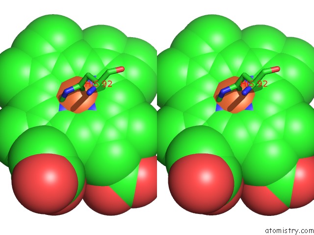
Iron binding site 2 out of 4 in 5u3i
Go back to
Iron binding site 2 out
of 4 in the Crystal Structure of Carbonmonoxy Hemoglobin S (Liganded Sickle Cell Hemoglobin) Complexed with Gbt Compound 31
Mono view
Stereo pair view
Mono view
Stereo pair view
A full contact list of Iron with other atoms in the Fe binding
site number 2 of Crystal Structure of Carbonmonoxy Hemoglobin S (Liganded Sickle Cell Hemoglobin) Complexed with Gbt Compound 31 within 5.0Å range:
|
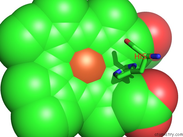
Iron binding site 3 out of 4 in 5u3i
Go back to
Iron binding site 3 out
of 4 in the Crystal Structure of Carbonmonoxy Hemoglobin S (Liganded Sickle Cell Hemoglobin) Complexed with Gbt Compound 31
Mono view
Stereo pair view
Mono view
Stereo pair view
A full contact list of Iron with other atoms in the Fe binding
site number 3 of Crystal Structure of Carbonmonoxy Hemoglobin S (Liganded Sickle Cell Hemoglobin) Complexed with Gbt Compound 31 within 5.0Å range:
|
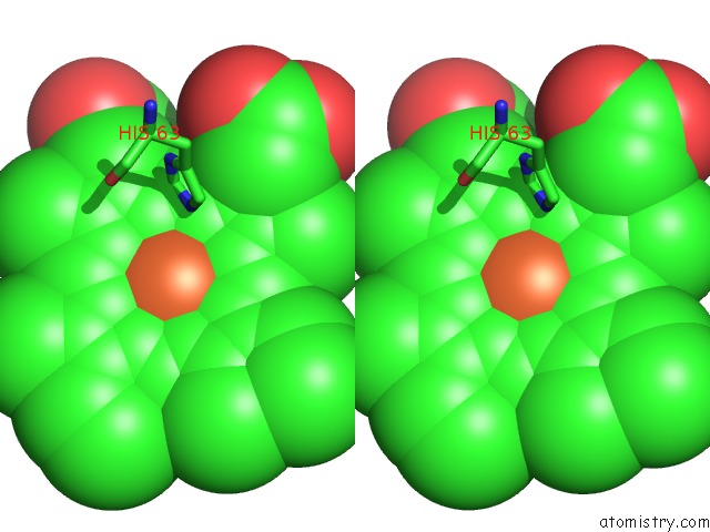
Iron binding site 4 out of 4 in 5u3i
Go back to
Iron binding site 4 out
of 4 in the Crystal Structure of Carbonmonoxy Hemoglobin S (Liganded Sickle Cell Hemoglobin) Complexed with Gbt Compound 31
Mono view
Stereo pair view
Mono view
Stereo pair view
A full contact list of Iron with other atoms in the Fe binding
site number 4 of Crystal Structure of Carbonmonoxy Hemoglobin S (Liganded Sickle Cell Hemoglobin) Complexed with Gbt Compound 31 within 5.0Å range:
|
Reference:
B.Metcalf,
C.Chuang,
K.Dufu,
M.P.Patel,
A.Silva-Garcia,
C.Johnson,
Q.Lu,
J.R.Partridge,
L.Patskovska,
Y.Patskovsky,
S.C.Almo,
M.P.Jacobson,
L.Hua,
Q.Xu,
S.L.Gwaltney,
C.Yee,
J.Harris,
B.P.Morgan,
J.James,
D.Xu,
A.Hutchaleelaha,
K.Paulvannan,
D.Oksenberg,
Z.Li.
Discovery of GBT440, An Orally Bioavailable R-State Stabilizer of Sickle Cell Hemoglobin. Acs Med Chem Lett V. 8 321 2017.
ISSN: ISSN 1948-5875
PubMed: 28337324
DOI: 10.1021/ACSMEDCHEMLETT.6B00491
Page generated: Tue Aug 6 09:44:20 2024
ISSN: ISSN 1948-5875
PubMed: 28337324
DOI: 10.1021/ACSMEDCHEMLETT.6B00491
Last articles
Zn in 9MJ5Zn in 9HNW
Zn in 9G0L
Zn in 9FNE
Zn in 9DZN
Zn in 9E0I
Zn in 9D32
Zn in 9DAK
Zn in 8ZXC
Zn in 8ZUF