Iron »
PDB 5tis-5ufj »
5u8x »
Iron in PDB 5u8x: Crystal Structure of Fe-CAO1
Protein crystallography data
The structure of Crystal Structure of Fe-CAO1, PDB code: 5u8x
was solved by
X.Sui,
K.Palczewski,
S.Banerjee,
P.D.Kiser,
with X-Ray Crystallography technique. A brief refinement statistics is given in the table below:
| Resolution Low / High (Å) | 50.14 / 2.17 |
| Space group | P 32 2 1 |
| Cell size a, b, c (Å), α, β, γ (°) | 100.908, 100.908, 448.815, 90.00, 90.00, 120.00 |
| R / Rfree (%) | 17.1 / 20.4 |
Other elements in 5u8x:
The structure of Crystal Structure of Fe-CAO1 also contains other interesting chemical elements:
| Chlorine | (Cl) | 4 atoms |
Iron Binding Sites:
The binding sites of Iron atom in the Crystal Structure of Fe-CAO1
(pdb code 5u8x). This binding sites where shown within
5.0 Angstroms radius around Iron atom.
In total 4 binding sites of Iron where determined in the Crystal Structure of Fe-CAO1, PDB code: 5u8x:
Jump to Iron binding site number: 1; 2; 3; 4;
In total 4 binding sites of Iron where determined in the Crystal Structure of Fe-CAO1, PDB code: 5u8x:
Jump to Iron binding site number: 1; 2; 3; 4;
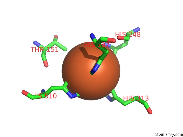
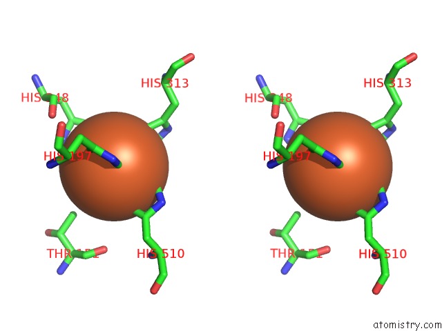
Iron binding site 1 out of 4 in 5u8x
Go back to
Iron binding site 1 out
of 4 in the Crystal Structure of Fe-CAO1
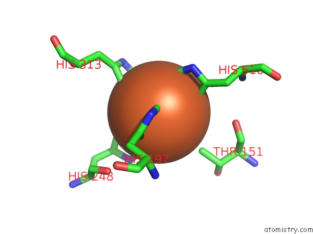
Mono view
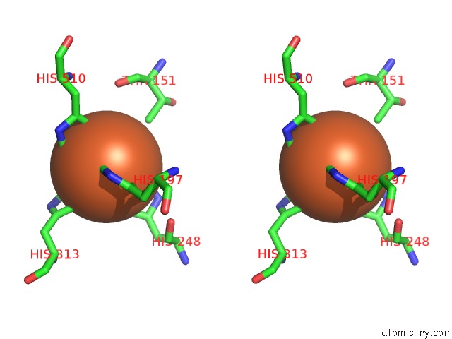
Stereo pair view
Mono view
Stereo pair view
A full contact list of Iron with other atoms in the Fe binding
site number 1 of Crystal Structure of Fe-CAO1 within 5.0Å range:
|
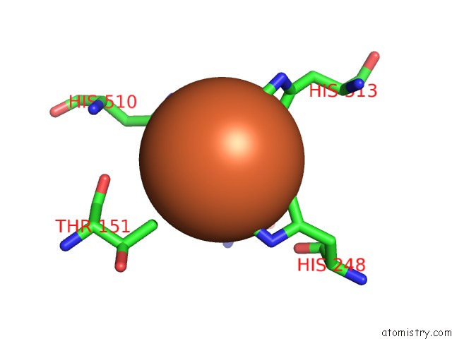
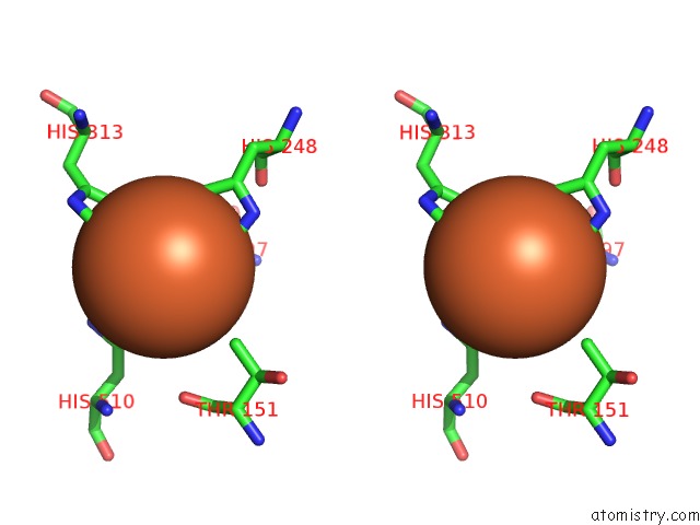
Iron binding site 2 out of 4 in 5u8x
Go back to
Iron binding site 2 out
of 4 in the Crystal Structure of Fe-CAO1
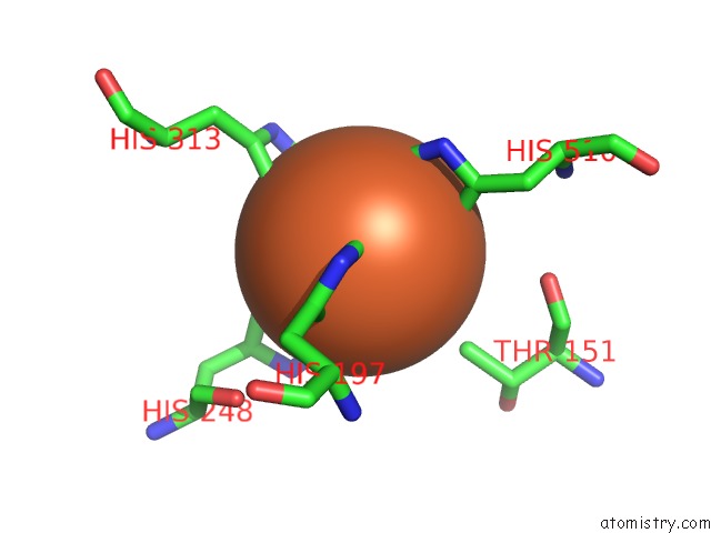
Mono view
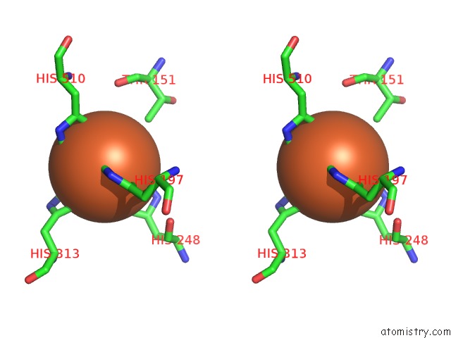
Stereo pair view
Mono view
Stereo pair view
A full contact list of Iron with other atoms in the Fe binding
site number 2 of Crystal Structure of Fe-CAO1 within 5.0Å range:
|
Iron binding site 3 out of 4 in 5u8x
Go back to
Iron binding site 3 out
of 4 in the Crystal Structure of Fe-CAO1
Mono view
Stereo pair view
Mono view
Stereo pair view
A full contact list of Iron with other atoms in the Fe binding
site number 3 of Crystal Structure of Fe-CAO1 within 5.0Å range:
|
Iron binding site 4 out of 4 in 5u8x
Go back to
Iron binding site 4 out
of 4 in the Crystal Structure of Fe-CAO1
Mono view
Stereo pair view
Mono view
Stereo pair view
A full contact list of Iron with other atoms in the Fe binding
site number 4 of Crystal Structure of Fe-CAO1 within 5.0Å range:
|
Reference:
X.Sui,
A.C.Weitz,
E.R.Farquhar,
M.Badiee,
S.Banerjee,
J.Von Lintig,
G.P.Tochtrop,
K.Palczewski,
M.P.Hendrich,
P.D.Kiser.
Structure and Spectroscopy of Alkene-Cleaving Dioxygenases Containing An Atypically Coordinated Non-Heme Iron Center. Biochemistry V. 56 2836 2017.
ISSN: ISSN 1520-4995
PubMed: 28493664
DOI: 10.1021/ACS.BIOCHEM.7B00251
Page generated: Wed Aug 6 01:50:42 2025
ISSN: ISSN 1520-4995
PubMed: 28493664
DOI: 10.1021/ACS.BIOCHEM.7B00251
Last articles
Fe in 6ZKLFe in 6ZKI
Fe in 6ZKK
Fe in 6ZKJ
Fe in 6ZKH
Fe in 6ZKG
Fe in 6ZKF
Fe in 6ZKE
Fe in 6ZKD
Fe in 6ZKC