Iron »
PDB 5utd-5ve3 »
5v1t »
Iron in PDB 5v1t: Crystal Structure of Streptococcus Suis Suib Bound to Precursor Peptide Suia
Protein crystallography data
The structure of Crystal Structure of Streptococcus Suis Suib Bound to Precursor Peptide Suia, PDB code: 5v1t
was solved by
K.M.Davis,
J.P.Bacik,
N.Ando,
with X-Ray Crystallography technique. A brief refinement statistics is given in the table below:
| Resolution Low / High (Å) | 29.24 / 2.10 |
| Space group | P 21 21 2 |
| Cell size a, b, c (Å), α, β, γ (°) | 69.370, 115.040, 54.330, 90.00, 90.00, 90.00 |
| R / Rfree (%) | 18.9 / 22.3 |
Iron Binding Sites:
Pages:
>>> Page 1 <<< Page 2, Binding sites: 11 - 12;Binding sites:
The binding sites of Iron atom in the Crystal Structure of Streptococcus Suis Suib Bound to Precursor Peptide Suia (pdb code 5v1t). This binding sites where shown within 5.0 Angstroms radius around Iron atom.In total 12 binding sites of Iron where determined in the Crystal Structure of Streptococcus Suis Suib Bound to Precursor Peptide Suia, PDB code: 5v1t:
Jump to Iron binding site number: 1; 2; 3; 4; 5; 6; 7; 8; 9; 10;
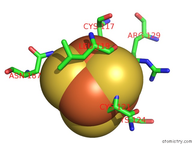
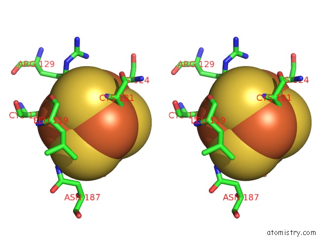
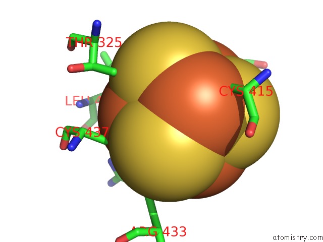
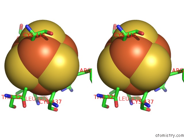
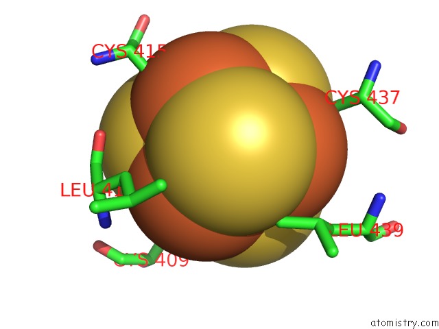
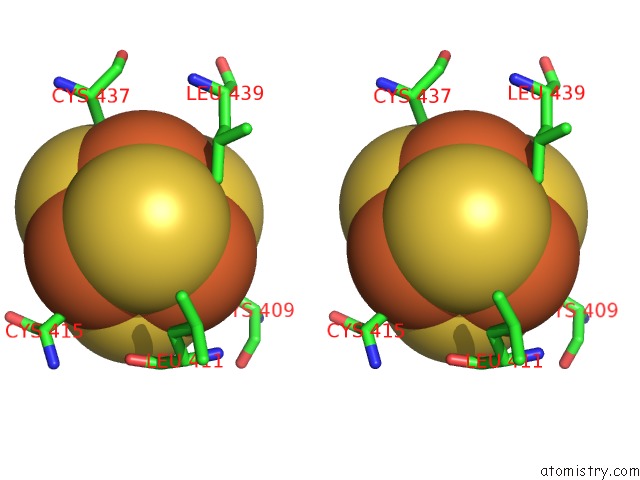
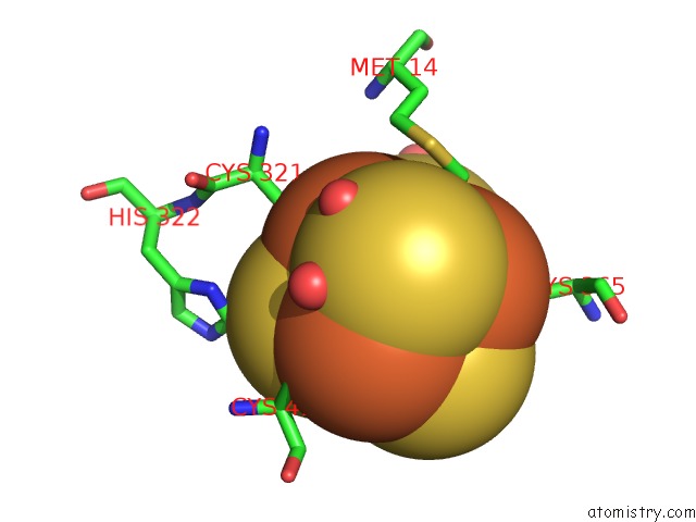
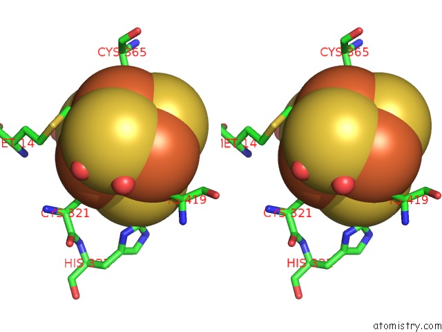
Iron binding site 1 out of 12 in 5v1t
Go back to
Iron binding site 1 out
of 12 in the Crystal Structure of Streptococcus Suis Suib Bound to Precursor Peptide Suia
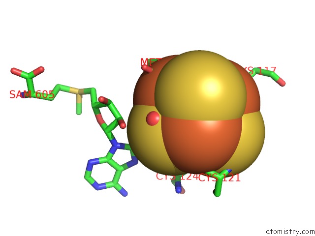
Mono view
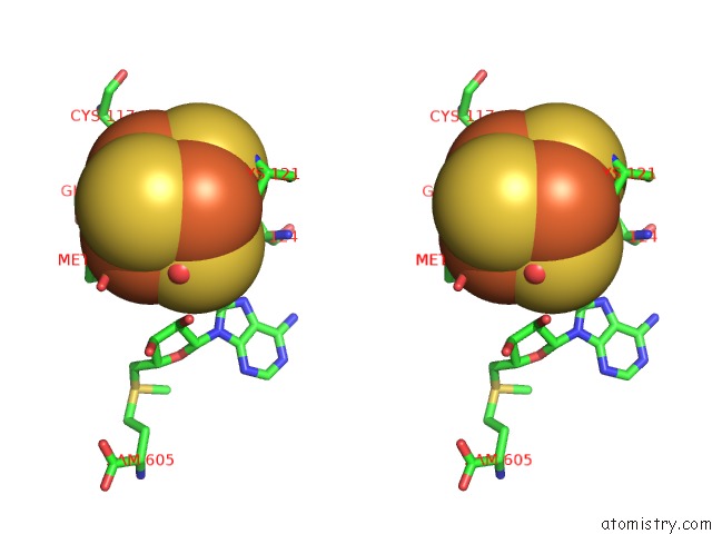
Stereo pair view
Mono view
Stereo pair view
A full contact list of Iron with other atoms in the Fe binding
site number 1 of Crystal Structure of Streptococcus Suis Suib Bound to Precursor Peptide Suia within 5.0Å range:
|
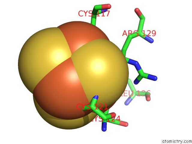
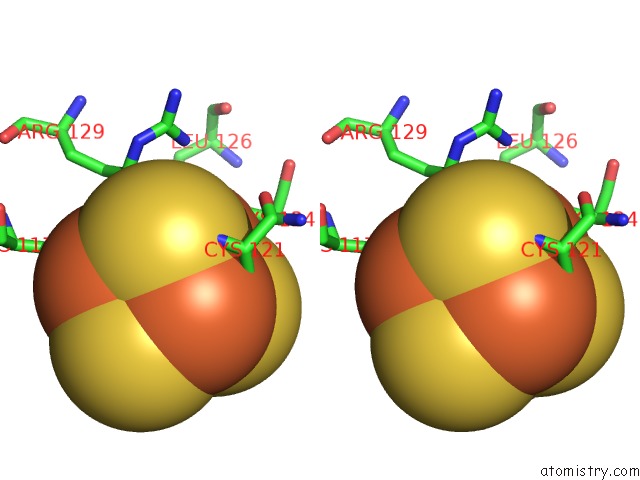
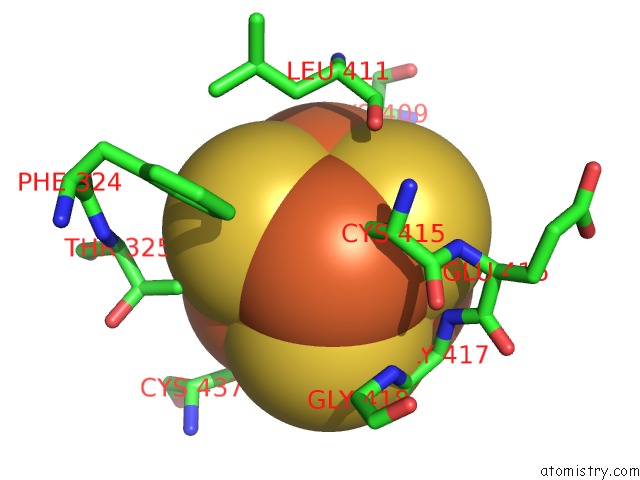
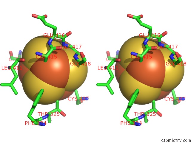
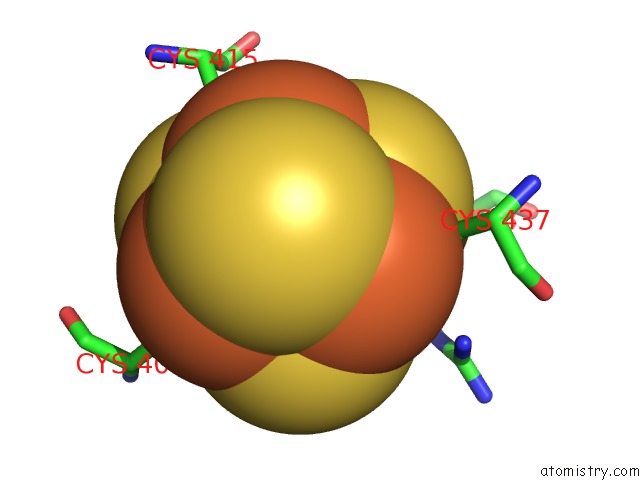
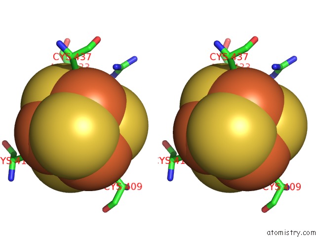
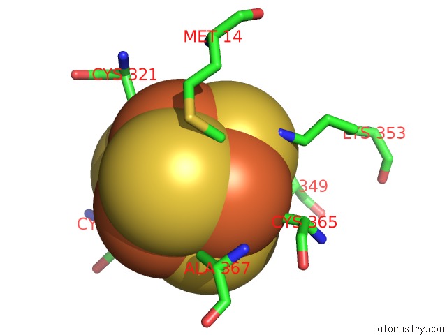
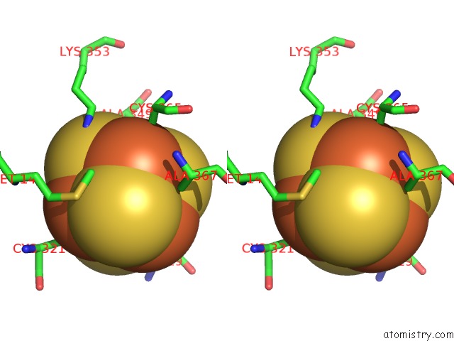
Iron binding site 2 out of 12 in 5v1t
Go back to
Iron binding site 2 out
of 12 in the Crystal Structure of Streptococcus Suis Suib Bound to Precursor Peptide Suia
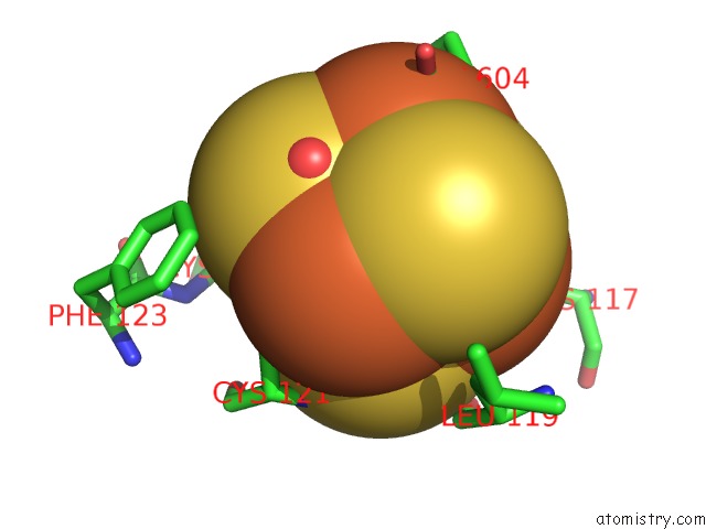
Mono view
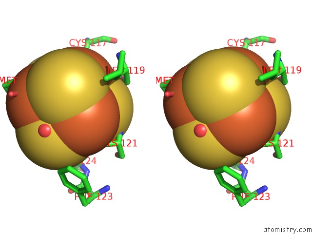
Stereo pair view
Mono view
Stereo pair view
A full contact list of Iron with other atoms in the Fe binding
site number 2 of Crystal Structure of Streptococcus Suis Suib Bound to Precursor Peptide Suia within 5.0Å range:
|
Iron binding site 3 out of 12 in 5v1t
Go back to
Iron binding site 3 out
of 12 in the Crystal Structure of Streptococcus Suis Suib Bound to Precursor Peptide Suia
Mono view
Stereo pair view
Mono view
Stereo pair view
A full contact list of Iron with other atoms in the Fe binding
site number 3 of Crystal Structure of Streptococcus Suis Suib Bound to Precursor Peptide Suia within 5.0Å range:
|
Iron binding site 4 out of 12 in 5v1t
Go back to
Iron binding site 4 out
of 12 in the Crystal Structure of Streptococcus Suis Suib Bound to Precursor Peptide Suia
Mono view
Stereo pair view
Mono view
Stereo pair view
A full contact list of Iron with other atoms in the Fe binding
site number 4 of Crystal Structure of Streptococcus Suis Suib Bound to Precursor Peptide Suia within 5.0Å range:
|
Iron binding site 5 out of 12 in 5v1t
Go back to
Iron binding site 5 out
of 12 in the Crystal Structure of Streptococcus Suis Suib Bound to Precursor Peptide Suia
Mono view
Stereo pair view
Mono view
Stereo pair view
A full contact list of Iron with other atoms in the Fe binding
site number 5 of Crystal Structure of Streptococcus Suis Suib Bound to Precursor Peptide Suia within 5.0Å range:
|
Iron binding site 6 out of 12 in 5v1t
Go back to
Iron binding site 6 out
of 12 in the Crystal Structure of Streptococcus Suis Suib Bound to Precursor Peptide Suia
Mono view
Stereo pair view
Mono view
Stereo pair view
A full contact list of Iron with other atoms in the Fe binding
site number 6 of Crystal Structure of Streptococcus Suis Suib Bound to Precursor Peptide Suia within 5.0Å range:
|
Iron binding site 7 out of 12 in 5v1t
Go back to
Iron binding site 7 out
of 12 in the Crystal Structure of Streptococcus Suis Suib Bound to Precursor Peptide Suia
Mono view
Stereo pair view
Mono view
Stereo pair view
A full contact list of Iron with other atoms in the Fe binding
site number 7 of Crystal Structure of Streptococcus Suis Suib Bound to Precursor Peptide Suia within 5.0Å range:
|
Iron binding site 8 out of 12 in 5v1t
Go back to
Iron binding site 8 out
of 12 in the Crystal Structure of Streptococcus Suis Suib Bound to Precursor Peptide Suia
Mono view
Stereo pair view
Mono view
Stereo pair view
A full contact list of Iron with other atoms in the Fe binding
site number 8 of Crystal Structure of Streptococcus Suis Suib Bound to Precursor Peptide Suia within 5.0Å range:
|
Iron binding site 9 out of 12 in 5v1t
Go back to
Iron binding site 9 out
of 12 in the Crystal Structure of Streptococcus Suis Suib Bound to Precursor Peptide Suia
Mono view
Stereo pair view
Mono view
Stereo pair view
A full contact list of Iron with other atoms in the Fe binding
site number 9 of Crystal Structure of Streptococcus Suis Suib Bound to Precursor Peptide Suia within 5.0Å range:
|
Iron binding site 10 out of 12 in 5v1t
Go back to
Iron binding site 10 out
of 12 in the Crystal Structure of Streptococcus Suis Suib Bound to Precursor Peptide Suia
Mono view
Stereo pair view
Mono view
Stereo pair view
A full contact list of Iron with other atoms in the Fe binding
site number 10 of Crystal Structure of Streptococcus Suis Suib Bound to Precursor Peptide Suia within 5.0Å range:
|
Reference:
K.M.Davis,
K.R.Schramma,
W.A.Hansen,
J.P.Bacik,
S.D.Khare,
M.R.Seyedsayamdost,
N.Ando.
Structures of the Peptide-Modifying Radical Sam Enzyme Suib Elucidate the Basis of Substrate Recognition. Proc. Natl. Acad. Sci. V. 114 10420 2017U.S.A..
ISSN: ESSN 1091-6490
PubMed: 28893989
DOI: 10.1073/PNAS.1703663114
Page generated: Tue Aug 6 10:05:53 2024
ISSN: ESSN 1091-6490
PubMed: 28893989
DOI: 10.1073/PNAS.1703663114
Last articles
Zn in 9J0NZn in 9J0O
Zn in 9J0P
Zn in 9FJX
Zn in 9EKB
Zn in 9C0F
Zn in 9CAH
Zn in 9CH0
Zn in 9CH3
Zn in 9CH1