Iron in PDB 5vsm: Crystal Structure of Viperin with Bound [4FE-4S] Cluster, 5'- Deoxyadenosine, and L-Methionine
Protein crystallography data
The structure of Crystal Structure of Viperin with Bound [4FE-4S] Cluster, 5'- Deoxyadenosine, and L-Methionine, PDB code: 5vsm
was solved by
M.K.Fenwick,
Y.Li,
P.Cresswell,
Y.Modis,
S.E.Ealick,
with X-Ray Crystallography technique. A brief refinement statistics is given in the table below:
Resolution Low / High (Å)
|
45.53 /
1.70
|
Space group
|
P 21 21 21
|
Cell size a, b, c (Å), α, β, γ (°)
|
59.364,
73.264,
141.939,
90.00,
90.00,
90.00
|
R / Rfree (%)
|
15.9 /
19.5
|
Iron Binding Sites:
The binding sites of Iron atom in the Crystal Structure of Viperin with Bound [4FE-4S] Cluster, 5'- Deoxyadenosine, and L-Methionine
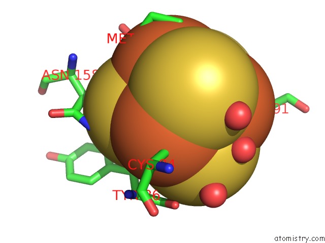
(pdb code 5vsm). This binding sites where shown within
5.0 Angstroms radius around Iron atom.
In total 8 binding sites of Iron where determined in the
Crystal Structure of Viperin with Bound [4FE-4S] Cluster, 5'- Deoxyadenosine, and L-Methionine, PDB code: 5vsm:
Jump to Iron binding site number:
1;
2;
3;
4;
5;
6;
7;
8;
Iron binding site 1 out
of 8 in 5vsm
Go back to
Iron Binding Sites List in 5vsm
Iron binding site 1 out
of 8 in the Crystal Structure of Viperin with Bound [4FE-4S] Cluster, 5'- Deoxyadenosine, and L-Methionine
 Mono view
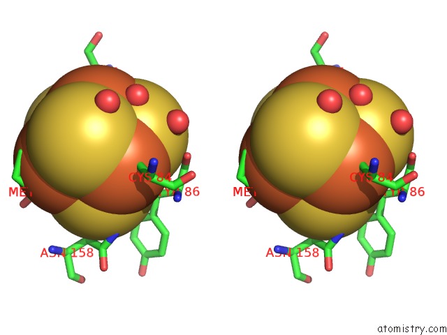
 Stereo pair view
|
A full contact list of Iron with other atoms in the Fe binding
site number 1 of Crystal Structure of Viperin with Bound [4FE-4S] Cluster, 5'- Deoxyadenosine, and L-Methionine within 5.0Å range:
probe
|
atom
|
residue
|
distance (Å)
|
B
|
Occ
|
A:Fe401
b:23.3
occ:1.00
|
FE1
|
A:SF4401
|
0.0
|
23.3
|
1.0
|
S2
|
A:SF4401
|
2.2
|
23.6
|
1.0
|
S4
|
A:SF4401
|
2.2
|
22.0
|
1.0
|
S3
|
A:SF4401
|
2.3
|
21.4
|
1.0
|
SG
|
A:CYS84
|
2.4
|
24.7
|
1.0
|
FE3
|
A:SF4401
|
2.6
|
23.5
|
1.0
|
FE2
|
A:SF4401
|
2.6
|
21.7
|
1.0
|
FE4
|
A:SF4401
|
2.8
|
24.2
|
1.0
|
CB
|
A:CYS84
|
3.3
|
24.0
|
1.0
|
S1
|
A:SF4401
|
3.8
|
23.6
|
1.0
|
N
|
A:MET403
|
3.8
|
22.5
|
1.0
|
CB
|
A:TYR86
|
4.0
|
25.6
|
1.0
|
O
|
A:HOH729
|
4.3
|
27.4
|
1.0
|
ND2
|
A:ASN158
|
4.6
|
20.2
|
1.0
|
O
|
A:TYR86
|
4.6
|
25.3
|
1.0
|
O
|
A:HOH532
|
4.6
|
26.9
|
1.0
|
N
|
A:TYR86
|
4.6
|
22.4
|
1.0
|
CA
|
A:TYR86
|
4.7
|
22.9
|
1.0
|
C
|
A:TYR86
|
4.7
|
24.8
|
1.0
|
CA
|
A:CYS84
|
4.7
|
28.3
|
1.0
|
O
|
A:MET403
|
4.7
|
25.4
|
1.0
|
SG
|
A:CYS91
|
4.8
|
23.8
|
1.0
|
O
|
A:HOH548
|
4.8
|
27.6
|
1.0
|
SG
|
A:CYS88
|
4.8
|
22.9
|
1.0
|
|
Iron binding site 2 out
of 8 in 5vsm
Go back to
Iron Binding Sites List in 5vsm
Iron binding site 2 out
of 8 in the Crystal Structure of Viperin with Bound [4FE-4S] Cluster, 5'- Deoxyadenosine, and L-Methionine
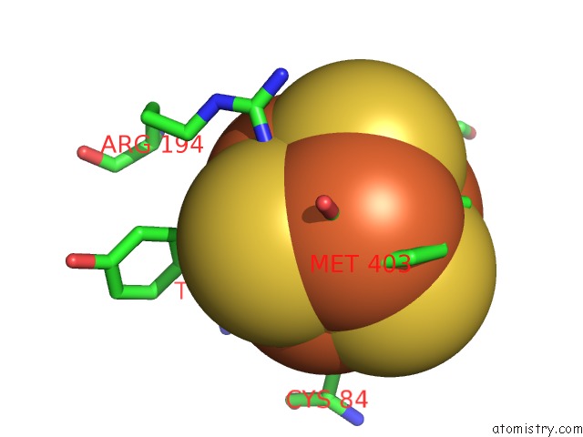
 Mono view
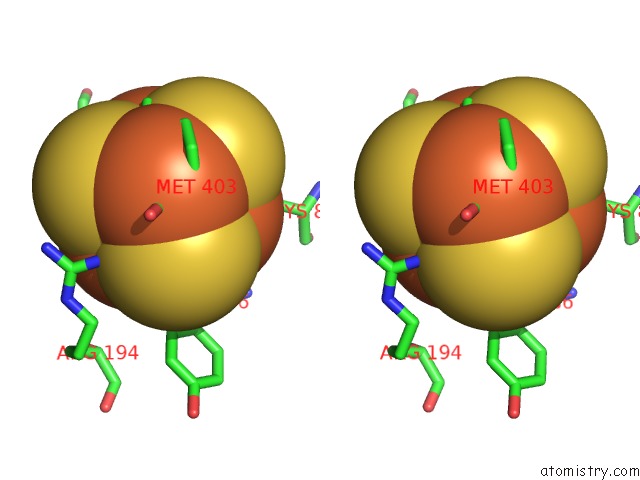
 Stereo pair view
|
A full contact list of Iron with other atoms in the Fe binding
site number 2 of Crystal Structure of Viperin with Bound [4FE-4S] Cluster, 5'- Deoxyadenosine, and L-Methionine within 5.0Å range:
probe
|
atom
|
residue
|
distance (Å)
|
B
|
Occ
|
A:Fe401
b:21.7
occ:1.00
|
FE2
|
A:SF4401
|
0.0
|
21.7
|
1.0
|
S1
|
A:SF4401
|
2.2
|
23.6
|
1.0
|
S3
|
A:SF4401
|
2.2
|
21.4
|
1.0
|
S4
|
A:SF4401
|
2.3
|
22.0
|
1.0
|
SG
|
A:CYS88
|
2.4
|
22.9
|
1.0
|
FE1
|
A:SF4401
|
2.6
|
23.3
|
1.0
|
FE3
|
A:SF4401
|
2.7
|
23.5
|
1.0
|
FE4
|
A:SF4401
|
2.9
|
24.2
|
1.0
|
CB
|
A:CYS88
|
3.1
|
22.6
|
1.0
|
NH1
|
A:ARG194
|
3.6
|
24.2
|
1.0
|
S2
|
A:SF4401
|
3.9
|
23.6
|
1.0
|
N
|
A:CYS88
|
4.0
|
23.2
|
1.0
|
CB
|
A:TYR86
|
4.1
|
25.6
|
1.0
|
O
|
A:MET403
|
4.2
|
25.4
|
1.0
|
CA
|
A:CYS88
|
4.2
|
23.5
|
1.0
|
CZ
|
A:ARG194
|
4.3
|
26.0
|
1.0
|
CB
|
A:CYS91
|
4.7
|
26.8
|
1.0
|
CD2
|
A:TYR86
|
4.7
|
24.3
|
1.0
|
SG
|
A:CYS84
|
4.7
|
24.7
|
1.0
|
SG
|
A:CYS91
|
4.8
|
23.8
|
1.0
|
NE
|
A:ARG194
|
4.8
|
27.5
|
1.0
|
CD
|
A:ARG194
|
4.9
|
26.1
|
1.0
|
NH2
|
A:ARG194
|
4.9
|
24.6
|
1.0
|
N
|
A:MET403
|
4.9
|
22.5
|
1.0
|
CG
|
A:TYR86
|
4.9
|
25.9
|
1.0
|
|
Iron binding site 3 out
of 8 in 5vsm
Go back to
Iron Binding Sites List in 5vsm
Iron binding site 3 out
of 8 in the Crystal Structure of Viperin with Bound [4FE-4S] Cluster, 5'- Deoxyadenosine, and L-Methionine
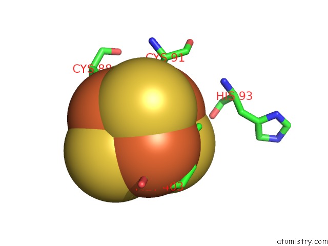
 Mono view
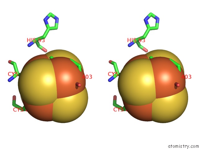
 Stereo pair view
|
A full contact list of Iron with other atoms in the Fe binding
site number 3 of Crystal Structure of Viperin with Bound [4FE-4S] Cluster, 5'- Deoxyadenosine, and L-Methionine within 5.0Å range:
probe
|
atom
|
residue
|
distance (Å)
|
B
|
Occ
|
A:Fe401
b:23.5
occ:1.00
|
FE3
|
A:SF4401
|
0.0
|
23.5
|
1.0
|
S1
|
A:SF4401
|
2.2
|
23.6
|
1.0
|
S2
|
A:SF4401
|
2.2
|
23.6
|
1.0
|
S4
|
A:SF4401
|
2.3
|
22.0
|
1.0
|
SG
|
A:CYS91
|
2.4
|
23.8
|
1.0
|
FE1
|
A:SF4401
|
2.6
|
23.3
|
1.0
|
FE2
|
A:SF4401
|
2.7
|
21.7
|
1.0
|
FE4
|
A:SF4401
|
2.8
|
24.2
|
1.0
|
CB
|
A:CYS91
|
3.1
|
26.8
|
1.0
|
O
|
A:HOH729
|
3.7
|
27.4
|
1.0
|
CE
|
A:MET403
|
3.8
|
24.2
|
1.0
|
S3
|
A:SF4401
|
3.9
|
21.4
|
1.0
|
CA
|
A:CYS91
|
4.1
|
24.0
|
1.0
|
SD
|
A:MET403
|
4.2
|
29.1
|
1.0
|
O
|
A:HOH548
|
4.5
|
27.6
|
1.0
|
O
|
A:HIS93
|
4.6
|
27.3
|
1.0
|
CB
|
A:CYS88
|
4.6
|
22.6
|
1.0
|
SG
|
A:CYS88
|
4.7
|
22.9
|
1.0
|
N
|
A:MET403
|
4.8
|
22.5
|
1.0
|
SG
|
A:CYS84
|
4.9
|
24.7
|
1.0
|
O
|
A:MET403
|
4.9
|
25.4
|
1.0
|
C
|
A:CYS91
|
4.9
|
28.8
|
1.0
|
|
Iron binding site 4 out
of 8 in 5vsm
Go back to
Iron Binding Sites List in 5vsm
Iron binding site 4 out
of 8 in the Crystal Structure of Viperin with Bound [4FE-4S] Cluster, 5'- Deoxyadenosine, and L-Methionine
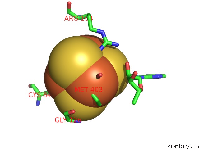
 Mono view
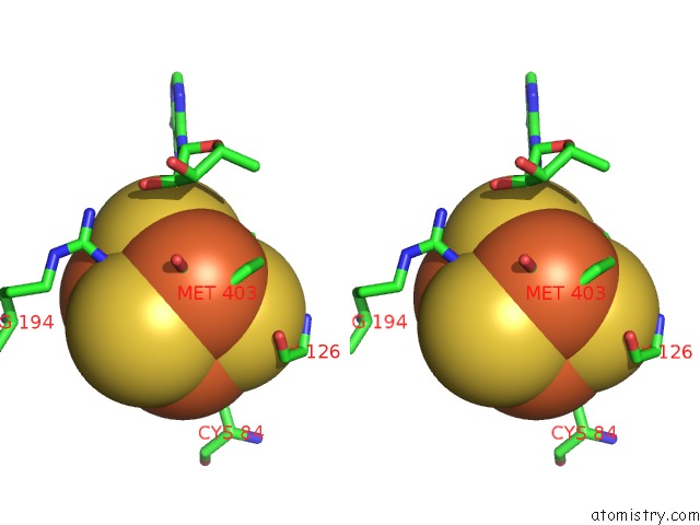
 Stereo pair view
|
A full contact list of Iron with other atoms in the Fe binding
site number 4 of Crystal Structure of Viperin with Bound [4FE-4S] Cluster, 5'- Deoxyadenosine, and L-Methionine within 5.0Å range:
probe
|
atom
|
residue
|
distance (Å)
|
B
|
Occ
|
A:Fe401
b:24.2
occ:1.00
|
FE4
|
A:SF4401
|
0.0
|
24.2
|
1.0
|
O
|
A:MET403
|
2.3
|
25.4
|
1.0
|
N
|
A:MET403
|
2.4
|
22.5
|
1.0
|
S2
|
A:SF4401
|
2.4
|
23.6
|
1.0
|
S1
|
A:SF4401
|
2.4
|
23.6
|
1.0
|
S3
|
A:SF4401
|
2.4
|
21.4
|
1.0
|
SD
|
A:MET403
|
2.7
|
29.1
|
1.0
|
FE1
|
A:SF4401
|
2.8
|
23.3
|
1.0
|
FE3
|
A:SF4401
|
2.8
|
23.5
|
1.0
|
FE2
|
A:SF4401
|
2.9
|
21.7
|
1.0
|
C
|
A:MET403
|
3.0
|
26.7
|
1.0
|
CA
|
A:MET403
|
3.1
|
23.9
|
1.0
|
CG
|
A:MET403
|
3.5
|
29.1
|
1.0
|
CE
|
A:MET403
|
3.7
|
24.2
|
1.0
|
CB
|
A:MET403
|
3.8
|
25.2
|
1.0
|
NH1
|
A:ARG194
|
3.9
|
24.2
|
1.0
|
S4
|
A:SF4401
|
4.1
|
22.0
|
1.0
|
OXT
|
A:MET403
|
4.3
|
23.7
|
1.0
|
O
|
A:GLY126
|
4.6
|
25.6
|
1.0
|
SG
|
A:CYS84
|
4.7
|
24.7
|
1.0
|
C2'
|
A:5AD402
|
4.8
|
26.9
|
1.0
|
SG
|
A:CYS91
|
5.0
|
23.8
|
1.0
|
|
Iron binding site 5 out
of 8 in 5vsm
Go back to
Iron Binding Sites List in 5vsm
Iron binding site 5 out
of 8 in the Crystal Structure of Viperin with Bound [4FE-4S] Cluster, 5'- Deoxyadenosine, and L-Methionine
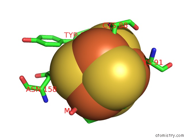
 Mono view
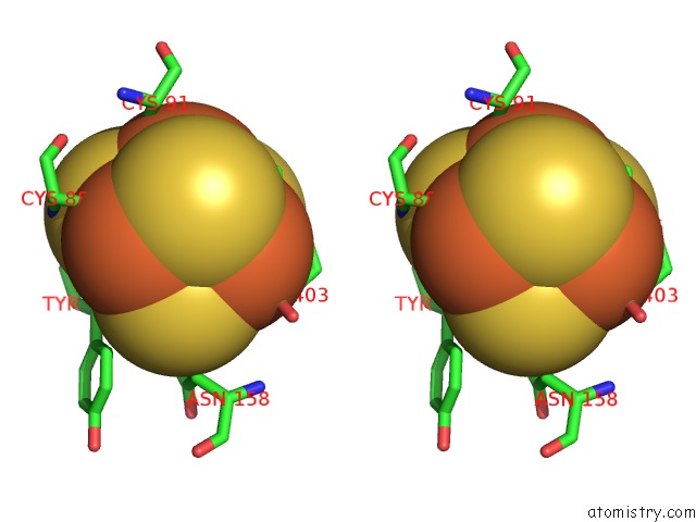
 Stereo pair view
|
A full contact list of Iron with other atoms in the Fe binding
site number 5 of Crystal Structure of Viperin with Bound [4FE-4S] Cluster, 5'- Deoxyadenosine, and L-Methionine within 5.0Å range:
probe
|
atom
|
residue
|
distance (Å)
|
B
|
Occ
|
B:Fe401
b:23.3
occ:1.00
|
FE1
|
B:SF4401
|
0.0
|
23.3
|
1.0
|
S2
|
B:SF4401
|
2.2
|
22.4
|
1.0
|
S4
|
B:SF4401
|
2.3
|
21.8
|
1.0
|
S3
|
B:SF4401
|
2.3
|
21.0
|
1.0
|
SG
|
B:CYS84
|
2.4
|
27.2
|
1.0
|
FE3
|
B:SF4401
|
2.6
|
24.9
|
1.0
|
FE2
|
B:SF4401
|
2.6
|
22.8
|
1.0
|
FE4
|
B:SF4401
|
2.7
|
24.0
|
1.0
|
CB
|
B:CYS84
|
3.3
|
28.8
|
1.0
|
S1
|
B:SF4401
|
3.7
|
23.5
|
1.0
|
N
|
B:MET403
|
3.8
|
23.2
|
1.0
|
CB
|
B:TYR86
|
4.0
|
23.6
|
1.0
|
O
|
B:HOH737
|
4.3
|
28.4
|
1.0
|
O
|
B:HOH558
|
4.5
|
29.3
|
1.0
|
O
|
B:TYR86
|
4.6
|
28.6
|
1.0
|
ND2
|
B:ASN158
|
4.6
|
23.2
|
1.0
|
N
|
B:TYR86
|
4.7
|
25.5
|
1.0
|
C
|
B:TYR86
|
4.7
|
27.7
|
1.0
|
CA
|
B:TYR86
|
4.7
|
24.3
|
1.0
|
CA
|
B:CYS84
|
4.8
|
28.2
|
1.0
|
O
|
B:MET403
|
4.8
|
22.4
|
1.0
|
SG
|
B:CYS88
|
4.8
|
24.6
|
1.0
|
SG
|
B:CYS91
|
4.9
|
28.9
|
1.0
|
O
|
B:HOH543
|
4.9
|
25.7
|
1.0
|
C
|
B:GLY126
|
5.0
|
26.1
|
1.0
|
|
Iron binding site 6 out
of 8 in 5vsm
Go back to
Iron Binding Sites List in 5vsm
Iron binding site 6 out
of 8 in the Crystal Structure of Viperin with Bound [4FE-4S] Cluster, 5'- Deoxyadenosine, and L-Methionine
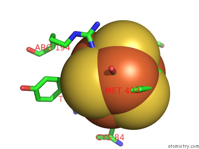
 Mono view
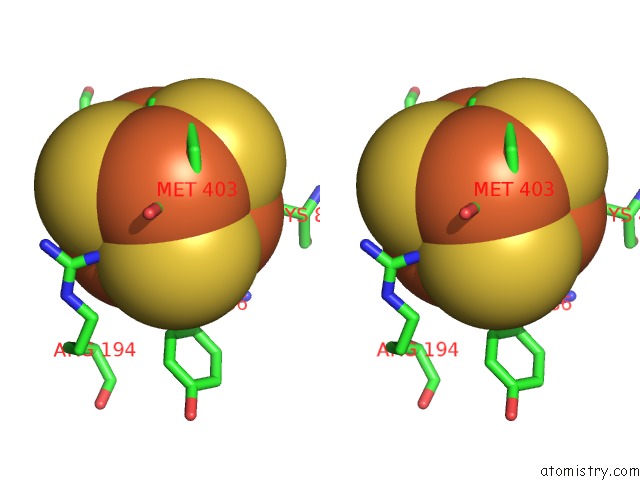
 Stereo pair view
|
A full contact list of Iron with other atoms in the Fe binding
site number 6 of Crystal Structure of Viperin with Bound [4FE-4S] Cluster, 5'- Deoxyadenosine, and L-Methionine within 5.0Å range:
probe
|
atom
|
residue
|
distance (Å)
|
B
|
Occ
|
B:Fe401
b:22.8
occ:1.00
|
FE2
|
B:SF4401
|
0.0
|
22.8
|
1.0
|
S1
|
B:SF4401
|
2.2
|
23.5
|
1.0
|
S3
|
B:SF4401
|
2.2
|
21.0
|
1.0
|
S4
|
B:SF4401
|
2.3
|
21.8
|
1.0
|
SG
|
B:CYS88
|
2.4
|
24.6
|
1.0
|
FE3
|
B:SF4401
|
2.6
|
24.9
|
1.0
|
FE1
|
B:SF4401
|
2.6
|
23.3
|
1.0
|
FE4
|
B:SF4401
|
2.8
|
24.0
|
1.0
|
CB
|
B:CYS88
|
3.2
|
23.2
|
1.0
|
NH1
|
B:ARG194
|
3.6
|
23.7
|
1.0
|
S2
|
B:SF4401
|
3.8
|
22.4
|
1.0
|
N
|
B:CYS88
|
4.1
|
23.7
|
1.0
|
CB
|
B:TYR86
|
4.1
|
23.6
|
1.0
|
O
|
B:MET403
|
4.2
|
22.4
|
1.0
|
CA
|
B:CYS88
|
4.2
|
22.4
|
1.0
|
CZ
|
B:ARG194
|
4.3
|
20.8
|
1.0
|
CB
|
B:CYS91
|
4.7
|
29.3
|
1.0
|
SG
|
B:CYS84
|
4.7
|
27.2
|
1.0
|
CD
|
B:ARG194
|
4.7
|
21.2
|
1.0
|
NE
|
B:ARG194
|
4.8
|
20.3
|
1.0
|
CD2
|
B:TYR86
|
4.8
|
24.5
|
1.0
|
SG
|
B:CYS91
|
4.8
|
28.9
|
1.0
|
NH2
|
B:ARG194
|
4.9
|
26.1
|
1.0
|
N
|
B:MET403
|
4.9
|
23.2
|
1.0
|
CG
|
B:TYR86
|
5.0
|
26.1
|
1.0
|
|
Iron binding site 7 out
of 8 in 5vsm
Go back to
Iron Binding Sites List in 5vsm
Iron binding site 7 out
of 8 in the Crystal Structure of Viperin with Bound [4FE-4S] Cluster, 5'- Deoxyadenosine, and L-Methionine
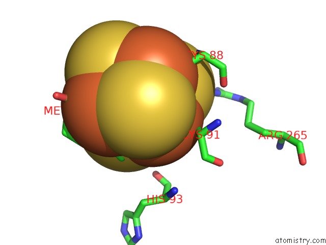
 Mono view
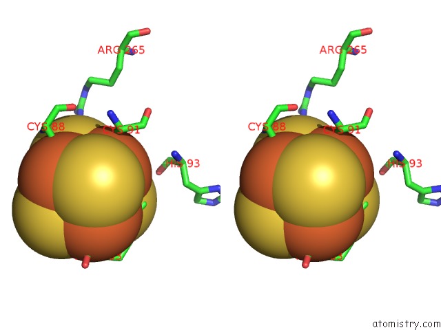
 Stereo pair view
|
A full contact list of Iron with other atoms in the Fe binding
site number 7 of Crystal Structure of Viperin with Bound [4FE-4S] Cluster, 5'- Deoxyadenosine, and L-Methionine within 5.0Å range:
probe
|
atom
|
residue
|
distance (Å)
|
B
|
Occ
|
B:Fe401
b:24.9
occ:1.00
|
FE3
|
B:SF4401
|
0.0
|
24.9
|
1.0
|
S2
|
B:SF4401
|
2.2
|
22.4
|
1.0
|
S1
|
B:SF4401
|
2.2
|
23.5
|
1.0
|
S4
|
B:SF4401
|
2.3
|
21.8
|
1.0
|
SG
|
B:CYS91
|
2.5
|
28.9
|
1.0
|
FE1
|
B:SF4401
|
2.6
|
23.3
|
1.0
|
FE2
|
B:SF4401
|
2.6
|
22.8
|
1.0
|
FE4
|
B:SF4401
|
2.8
|
24.0
|
1.0
|
CB
|
B:CYS91
|
3.2
|
29.3
|
1.0
|
CE
|
B:MET403
|
3.8
|
27.4
|
1.0
|
S3
|
B:SF4401
|
3.8
|
21.0
|
1.0
|
O
|
B:HOH737
|
3.8
|
28.4
|
1.0
|
CA
|
B:CYS91
|
4.1
|
28.0
|
1.0
|
SD
|
B:MET403
|
4.1
|
27.5
|
1.0
|
O
|
B:HOH543
|
4.6
|
25.7
|
1.0
|
CB
|
B:CYS88
|
4.6
|
23.2
|
1.0
|
O
|
B:HIS93
|
4.7
|
27.7
|
1.0
|
SG
|
B:CYS88
|
4.8
|
24.6
|
1.0
|
SG
|
B:CYS84
|
4.9
|
27.2
|
1.0
|
N
|
B:MET403
|
4.9
|
23.2
|
1.0
|
O
|
B:MET403
|
5.0
|
22.4
|
1.0
|
NH1
|
B:ARG265
|
5.0
|
33.4
|
1.0
|
C
|
B:CYS91
|
5.0
|
29.8
|
1.0
|
|
Iron binding site 8 out
of 8 in 5vsm
Go back to
Iron Binding Sites List in 5vsm
Iron binding site 8 out
of 8 in the Crystal Structure of Viperin with Bound [4FE-4S] Cluster, 5'- Deoxyadenosine, and L-Methionine
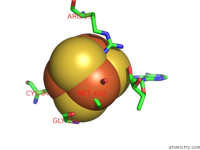
 Mono view
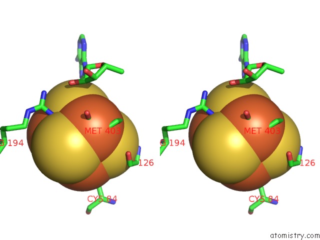
 Stereo pair view
|
A full contact list of Iron with other atoms in the Fe binding
site number 8 of Crystal Structure of Viperin with Bound [4FE-4S] Cluster, 5'- Deoxyadenosine, and L-Methionine within 5.0Å range:
probe
|
atom
|
residue
|
distance (Å)
|
B
|
Occ
|
B:Fe401
b:24.0
occ:1.00
|
FE4
|
B:SF4401
|
0.0
|
24.0
|
1.0
|
S3
|
B:SF4401
|
2.3
|
21.0
|
1.0
|
O
|
B:MET403
|
2.3
|
22.4
|
1.0
|
S2
|
B:SF4401
|
2.4
|
22.4
|
1.0
|
S1
|
B:SF4401
|
2.4
|
23.5
|
1.0
|
N
|
B:MET403
|
2.4
|
23.2
|
1.0
|
FE1
|
B:SF4401
|
2.7
|
23.3
|
1.0
|
SD
|
B:MET403
|
2.7
|
27.5
|
1.0
|
FE3
|
B:SF4401
|
2.8
|
24.9
|
1.0
|
FE2
|
B:SF4401
|
2.8
|
22.8
|
1.0
|
C
|
B:MET403
|
3.0
|
32.4
|
1.0
|
CA
|
B:MET403
|
3.2
|
27.3
|
1.0
|
CG
|
B:MET403
|
3.4
|
25.4
|
1.0
|
CE
|
B:MET403
|
3.7
|
27.4
|
1.0
|
CB
|
B:MET403
|
3.8
|
26.0
|
1.0
|
NH1
|
B:ARG194
|
3.9
|
23.7
|
1.0
|
S4
|
B:SF4401
|
4.1
|
21.8
|
1.0
|
OXT
|
B:MET403
|
4.3
|
24.6
|
1.0
|
O
|
B:GLY126
|
4.6
|
25.4
|
1.0
|
SG
|
B:CYS84
|
4.6
|
27.2
|
1.0
|
C2'
|
B:5AD402
|
4.8
|
25.3
|
1.0
|
O2'
|
B:5AD402
|
4.9
|
27.1
|
1.0
|
|
Reference:
M.K.Fenwick,
Y.Li,
P.Cresswell,
Y.Modis,
S.E.Ealick.
Structural Studies of Viperin, An Antiviral Radical Sam Enzyme. Proc. Natl. Acad. Sci. V. 114 6806 2017U.S.A..
ISSN: ESSN 1091-6490
PubMed: 28607080
DOI: 10.1073/PNAS.1705402114
Page generated: Tue Aug 6 10:33:44 2024
|