Iron »
PDB 5why-5xbp »
5wqr »
Iron in PDB 5wqr: High Resolution Structure of High-Potential Iron-Sulfur Protein in the Reduced State
Protein crystallography data
The structure of High Resolution Structure of High-Potential Iron-Sulfur Protein in the Reduced State, PDB code: 5wqr
was solved by
H.Ohno,
K.Takeda,
S.Niwa,
T.Tsujinaka,
Y.Hanazono,
Y.Hirano,
K.Miki,
with X-Ray Crystallography technique. A brief refinement statistics is given in the table below:
| Resolution Low / High (Å) | 14.96 / 0.80 |
| Space group | P 21 21 21 |
| Cell size a, b, c (Å), α, β, γ (°) | 46.330, 58.811, 23.423, 90.00, 90.00, 90.00 |
| R / Rfree (%) | 10.6 / 11.8 |
Iron Binding Sites:
The binding sites of Iron atom in the High Resolution Structure of High-Potential Iron-Sulfur Protein in the Reduced State
(pdb code 5wqr). This binding sites where shown within
5.0 Angstroms radius around Iron atom.
In total 4 binding sites of Iron where determined in the High Resolution Structure of High-Potential Iron-Sulfur Protein in the Reduced State, PDB code: 5wqr:
Jump to Iron binding site number: 1; 2; 3; 4;
In total 4 binding sites of Iron where determined in the High Resolution Structure of High-Potential Iron-Sulfur Protein in the Reduced State, PDB code: 5wqr:
Jump to Iron binding site number: 1; 2; 3; 4;
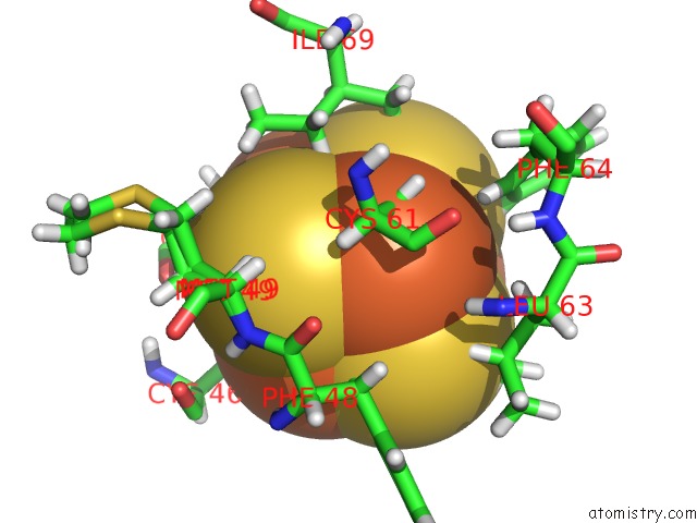
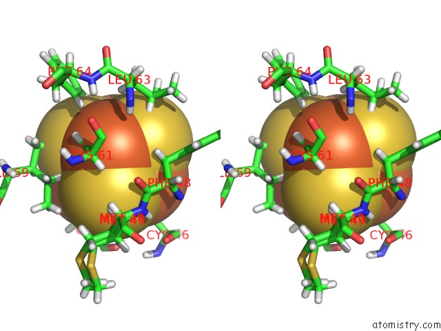
Iron binding site 1 out of 4 in 5wqr
Go back to
Iron binding site 1 out
of 4 in the High Resolution Structure of High-Potential Iron-Sulfur Protein in the Reduced State
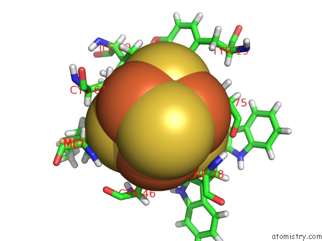
Mono view
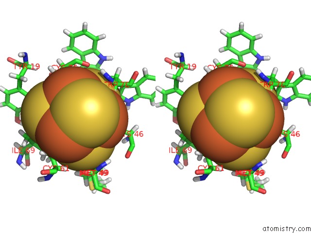
Stereo pair view
Mono view
Stereo pair view
A full contact list of Iron with other atoms in the Fe binding
site number 1 of High Resolution Structure of High-Potential Iron-Sulfur Protein in the Reduced State within 5.0Å range:
|
Iron binding site 2 out of 4 in 5wqr
Go back to
Iron binding site 2 out
of 4 in the High Resolution Structure of High-Potential Iron-Sulfur Protein in the Reduced State
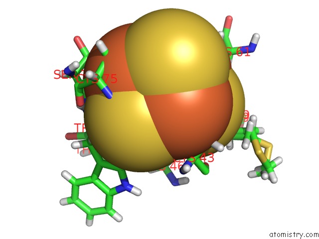
Mono view
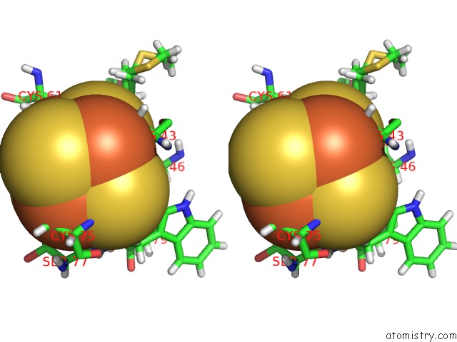
Stereo pair view
Mono view
Stereo pair view
A full contact list of Iron with other atoms in the Fe binding
site number 2 of High Resolution Structure of High-Potential Iron-Sulfur Protein in the Reduced State within 5.0Å range:
|
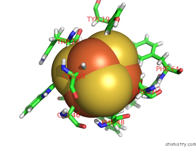
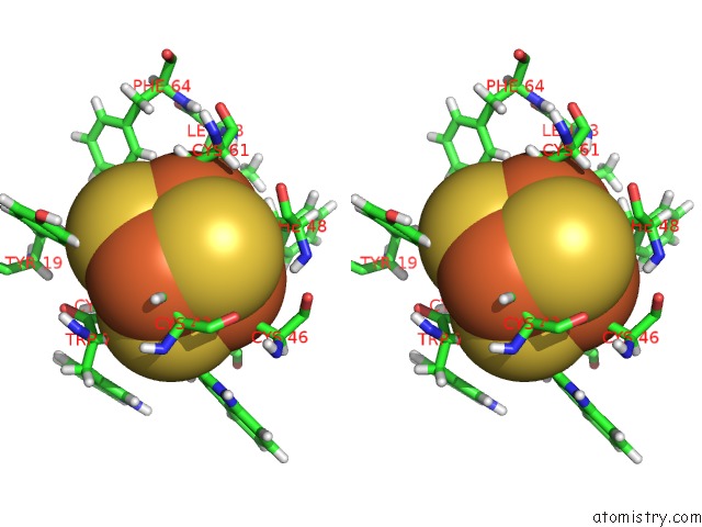
Iron binding site 3 out of 4 in 5wqr
Go back to
Iron binding site 3 out
of 4 in the High Resolution Structure of High-Potential Iron-Sulfur Protein in the Reduced State
Mono view
Stereo pair view
Mono view
Stereo pair view
A full contact list of Iron with other atoms in the Fe binding
site number 3 of High Resolution Structure of High-Potential Iron-Sulfur Protein in the Reduced State within 5.0Å range:
|
Iron binding site 4 out of 4 in 5wqr
Go back to
Iron binding site 4 out
of 4 in the High Resolution Structure of High-Potential Iron-Sulfur Protein in the Reduced State
Mono view
Stereo pair view
Mono view
Stereo pair view
A full contact list of Iron with other atoms in the Fe binding
site number 4 of High Resolution Structure of High-Potential Iron-Sulfur Protein in the Reduced State within 5.0Å range:
|
Reference:
H.Ohno,
K.Takeda,
S.Niwa,
T.Tsujinaka,
Y.Hanazono,
Y.Hirano,
K.Miki.
Crystallographic Characterization of the High-Potential Iron-Sulfur Protein in the Oxidized State at 0.8 Angstrom Resolution Plos One V. 12 78183 2017.
ISSN: ESSN 1932-6203
PubMed: 28542634
DOI: 10.1371/JOURNAL.PONE.0178183
Page generated: Wed Aug 6 02:33:46 2025
ISSN: ESSN 1932-6203
PubMed: 28542634
DOI: 10.1371/JOURNAL.PONE.0178183
Last articles
Fe in 6CDKFe in 6CII
Fe in 6CFW
Fe in 6CIF
Fe in 6CIE
Fe in 6CID
Fe in 6CIC
Fe in 6CHI
Fe in 6CI0
Fe in 6CH6